Iron »
PDB 101m-1a8f »
1a6g »
Iron in PDB 1a6g: Carbonmonoxy-Myoglobin, Atomic Resolution
Protein crystallography data
The structure of Carbonmonoxy-Myoglobin, Atomic Resolution, PDB code: 1a6g
was solved by
J.Vojtechovsky,
K.Chu,
J.Berendzen,
R.M.Sweet,
I.Schlichting,
with X-Ray Crystallography technique. A brief refinement statistics is given in the table below:
| Resolution Low / High (Å) | 8.00 / 1.15 |
| Space group | P 1 21 1 |
| Cell size a, b, c (Å), α, β, γ (°) | 63.800, 30.630, 34.420, 90.00, 105.80, 90.00 |
| R / Rfree (%) | 12.7 / 16 |
Iron Binding Sites:
The binding sites of Iron atom in the Carbonmonoxy-Myoglobin, Atomic Resolution
(pdb code 1a6g). This binding sites where shown within
5.0 Angstroms radius around Iron atom.
In total only one binding site of Iron was determined in the Carbonmonoxy-Myoglobin, Atomic Resolution, PDB code: 1a6g:
In total only one binding site of Iron was determined in the Carbonmonoxy-Myoglobin, Atomic Resolution, PDB code: 1a6g:
Iron binding site 1 out of 1 in 1a6g
Go back to
Iron binding site 1 out
of 1 in the Carbonmonoxy-Myoglobin, Atomic Resolution
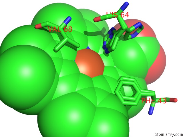
Mono view
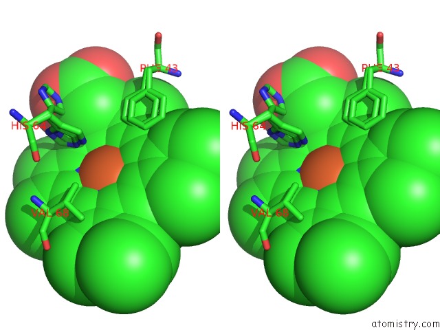
Stereo pair view
Mono view
Stereo pair view
A full contact list of Iron with other atoms in the Fe binding
site number 1 of Carbonmonoxy-Myoglobin, Atomic Resolution within 5.0Å range:
|
Reference:
J.Vojtechovsky,
K.Chu,
J.Berendzen,
R.M.Sweet,
I.Schlichting.
Crystal Structures of Myoglobin-Ligand Complexes at Near-Atomic Resolution. Biophys.J. V. 77 2153 1999.
ISSN: ISSN 0006-3495
PubMed: 10512835
Page generated: Wed Jul 16 12:02:58 2025
ISSN: ISSN 0006-3495
PubMed: 10512835
Last articles
Fe in 2YXOFe in 2YRS
Fe in 2YXC
Fe in 2YNM
Fe in 2YVJ
Fe in 2YP1
Fe in 2YU2
Fe in 2YU1
Fe in 2YQB
Fe in 2YOO