Iron »
PDB 1a9w-1aop »
1a9w »
Iron in PDB 1a9w: Human Embryonic Gower II Carbonmonoxy Hemoglobin
Protein crystallography data
The structure of Human Embryonic Gower II Carbonmonoxy Hemoglobin, PDB code: 1a9w
was solved by
A.J.Sutherland-Smith,
H.M.Baker,
O.M.Hofmann,
T.Brittain,
E.N.Baker,
with X-Ray Crystallography technique. A brief refinement statistics is given in the table below:
| Resolution Low / High (Å) | 40.00 / 2.90 |
| Space group | P 43 21 2 |
| Cell size a, b, c (Å), α, β, γ (°) | 62.800, 62.800, 320.900, 90.00, 90.00, 90.00 |
| R / Rfree (%) | 18.5 / 23.2 |
Iron Binding Sites:
The binding sites of Iron atom in the Human Embryonic Gower II Carbonmonoxy Hemoglobin
(pdb code 1a9w). This binding sites where shown within
5.0 Angstroms radius around Iron atom.
In total 4 binding sites of Iron where determined in the Human Embryonic Gower II Carbonmonoxy Hemoglobin, PDB code: 1a9w:
Jump to Iron binding site number: 1; 2; 3; 4;
In total 4 binding sites of Iron where determined in the Human Embryonic Gower II Carbonmonoxy Hemoglobin, PDB code: 1a9w:
Jump to Iron binding site number: 1; 2; 3; 4;
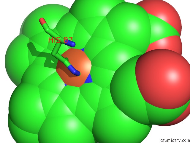
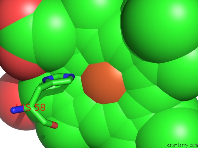
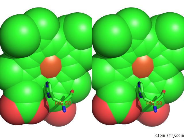
Iron binding site 1 out of 4 in 1a9w
Go back to
Iron binding site 1 out
of 4 in the Human Embryonic Gower II Carbonmonoxy Hemoglobin
Mono view
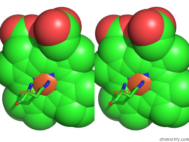
Stereo pair view
Mono view
Stereo pair view
A full contact list of Iron with other atoms in the Fe binding
site number 1 of Human Embryonic Gower II Carbonmonoxy Hemoglobin within 5.0Å range:
|
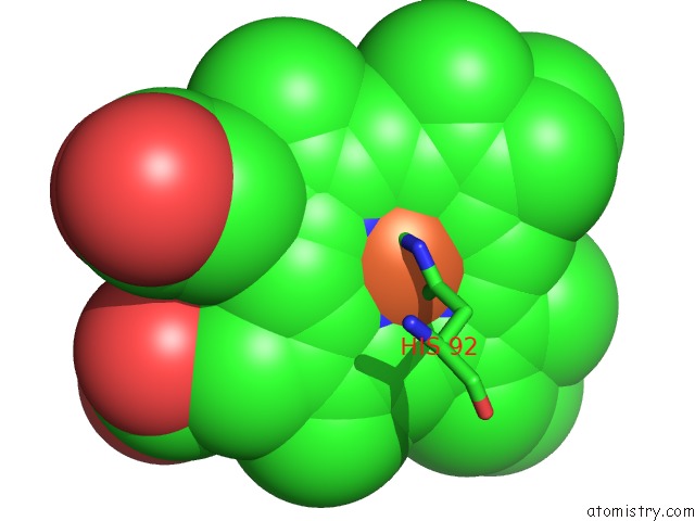
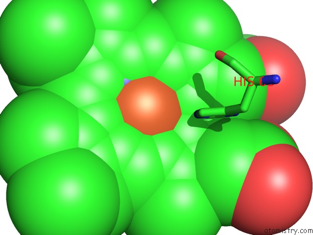
Iron binding site 2 out of 4 in 1a9w
Go back to
Iron binding site 2 out
of 4 in the Human Embryonic Gower II Carbonmonoxy Hemoglobin
Mono view
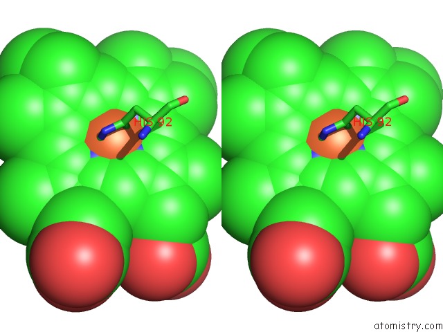
Stereo pair view
Mono view
Stereo pair view
A full contact list of Iron with other atoms in the Fe binding
site number 2 of Human Embryonic Gower II Carbonmonoxy Hemoglobin within 5.0Å range:
|
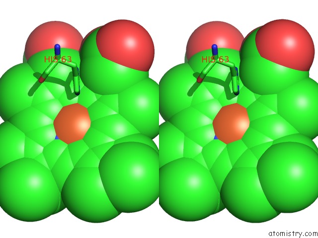
Iron binding site 3 out of 4 in 1a9w
Go back to
Iron binding site 3 out
of 4 in the Human Embryonic Gower II Carbonmonoxy Hemoglobin
Mono view
Stereo pair view
Mono view
Stereo pair view
A full contact list of Iron with other atoms in the Fe binding
site number 3 of Human Embryonic Gower II Carbonmonoxy Hemoglobin within 5.0Å range:
|
Iron binding site 4 out of 4 in 1a9w
Go back to
Iron binding site 4 out
of 4 in the Human Embryonic Gower II Carbonmonoxy Hemoglobin
Mono view
Stereo pair view
Mono view
Stereo pair view
A full contact list of Iron with other atoms in the Fe binding
site number 4 of Human Embryonic Gower II Carbonmonoxy Hemoglobin within 5.0Å range:
|
Reference:
A.J.Sutherland-Smith,
H.M.Baker,
O.M.Hofmann,
T.Brittain,
E.N.Baker.
Crystal Structure of A Human Embryonic Haemoglobin: the Carbonmonoxy Form of Gower II (ALPHA2 EPSILON2) Haemoglobin at 2.9 A Resolution. J.Mol.Biol. V. 280 475 1998.
ISSN: ISSN 0022-2836
PubMed: 9665850
DOI: 10.1006/JMBI.1998.1868
Page generated: Wed Jul 16 12:05:19 2025
ISSN: ISSN 0022-2836
PubMed: 9665850
DOI: 10.1006/JMBI.1998.1868
Last articles
Fe in 2YXOFe in 2YRS
Fe in 2YXC
Fe in 2YNM
Fe in 2YVJ
Fe in 2YP1
Fe in 2YU2
Fe in 2YU1
Fe in 2YQB
Fe in 2YOO