Iron »
PDB 1b2o-1biy »
1b2o »
Iron in PDB 1b2o: Clostridium Pasteurianum Rubredoxin G10VG43A Mutant
Protein crystallography data
The structure of Clostridium Pasteurianum Rubredoxin G10VG43A Mutant, PDB code: 1b2o
was solved by
M.J.Maher,
J.M.Guss,
M.C.J.Wilce,
A.G.Wedd,
with X-Ray Crystallography technique. A brief refinement statistics is given in the table below:
| Resolution Low / High (Å) | 30.00 / 1.90 |
| Space group | P 43 21 2 |
| Cell size a, b, c (Å), α, β, γ (°) | 61.850, 61.850, 80.450, 90.00, 90.00, 90.00 |
| R / Rfree (%) | 19.4 / 23.7 |
Iron Binding Sites:
The binding sites of Iron atom in the Clostridium Pasteurianum Rubredoxin G10VG43A Mutant
(pdb code 1b2o). This binding sites where shown within
5.0 Angstroms radius around Iron atom.
In total 2 binding sites of Iron where determined in the Clostridium Pasteurianum Rubredoxin G10VG43A Mutant, PDB code: 1b2o:
Jump to Iron binding site number: 1; 2;
In total 2 binding sites of Iron where determined in the Clostridium Pasteurianum Rubredoxin G10VG43A Mutant, PDB code: 1b2o:
Jump to Iron binding site number: 1; 2;
Iron binding site 1 out of 2 in 1b2o
Go back to
Iron binding site 1 out
of 2 in the Clostridium Pasteurianum Rubredoxin G10VG43A Mutant
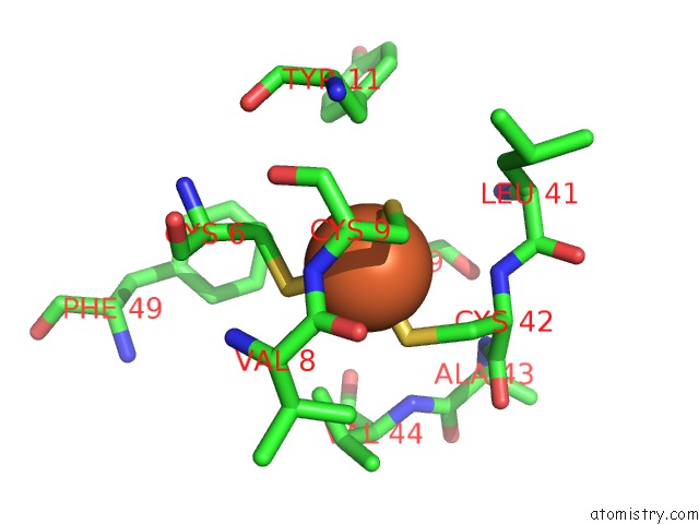
Mono view
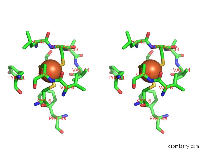
Stereo pair view
Mono view
Stereo pair view
A full contact list of Iron with other atoms in the Fe binding
site number 1 of Clostridium Pasteurianum Rubredoxin G10VG43A Mutant within 5.0Å range:
|
Iron binding site 2 out of 2 in 1b2o
Go back to
Iron binding site 2 out
of 2 in the Clostridium Pasteurianum Rubredoxin G10VG43A Mutant
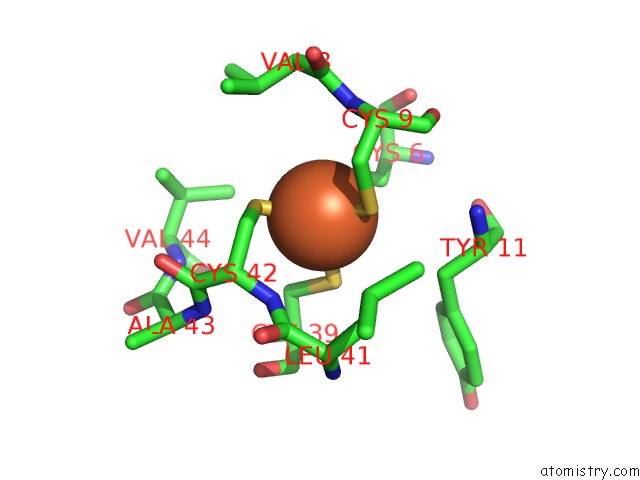
Mono view
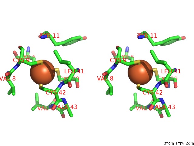
Stereo pair view
Mono view
Stereo pair view
A full contact list of Iron with other atoms in the Fe binding
site number 2 of Clostridium Pasteurianum Rubredoxin G10VG43A Mutant within 5.0Å range:
|
Reference:
M.J.Maher,
Z.Xiao,
M.C.Wilce,
J.M.Guss,
A.G.Wedd.
Rubredoxin From Clostridium Pasteurianum. Structures of G10A, G43A and G10VG43A Mutant Proteins. Mutation of Conserved Glycine 10 to Valine Causes the 9-10 Peptide Link to Invert. Acta Crystallogr.,Sect.D V. 55 962 1999.
ISSN: ISSN 0907-4449
PubMed: 10216292
DOI: 10.1107/S0907444999001900
Page generated: Sat Aug 3 02:39:00 2024
ISSN: ISSN 0907-4449
PubMed: 10216292
DOI: 10.1107/S0907444999001900
Last articles
Zn in 9MJ5Zn in 9HNW
Zn in 9G0L
Zn in 9FNE
Zn in 9DZN
Zn in 9E0I
Zn in 9D32
Zn in 9DAK
Zn in 8ZXC
Zn in 8ZUF