Iron »
PDB 1b2o-1biy »
1bin »
Iron in PDB 1bin: Leghemoglobin A (Acetomet)
Protein crystallography data
The structure of Leghemoglobin A (Acetomet), PDB code: 1bin
was solved by
E.A.Brucker,
M.S.Hargrove,
G.N.Phillips Jr.,
with X-Ray Crystallography technique. A brief refinement statistics is given in the table below:
| Resolution Low / High (Å) | 10.00 / 2.20 |
| Space group | P 21 21 21 |
| Cell size a, b, c (Å), α, β, γ (°) | 34.980, 53.390, 141.730, 90.00, 90.00, 90.00 |
| R / Rfree (%) | 19.8 / 29.7 |
Iron Binding Sites:
The binding sites of Iron atom in the Leghemoglobin A (Acetomet)
(pdb code 1bin). This binding sites where shown within
5.0 Angstroms radius around Iron atom.
In total 2 binding sites of Iron where determined in the Leghemoglobin A (Acetomet), PDB code: 1bin:
Jump to Iron binding site number: 1; 2;
In total 2 binding sites of Iron where determined in the Leghemoglobin A (Acetomet), PDB code: 1bin:
Jump to Iron binding site number: 1; 2;
Iron binding site 1 out of 2 in 1bin
Go back to
Iron binding site 1 out
of 2 in the Leghemoglobin A (Acetomet)
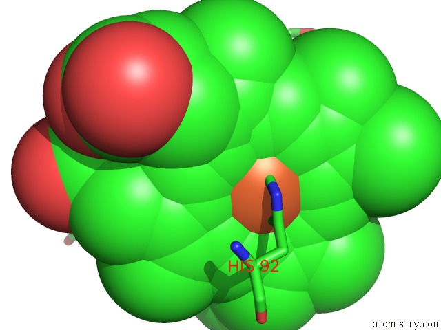
Mono view
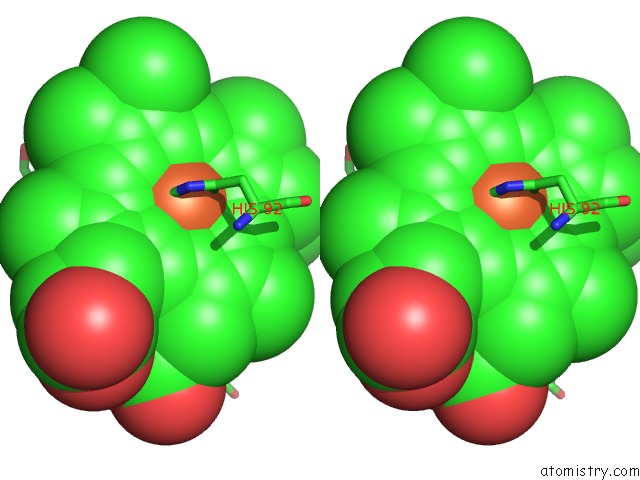
Stereo pair view
Mono view
Stereo pair view
A full contact list of Iron with other atoms in the Fe binding
site number 1 of Leghemoglobin A (Acetomet) within 5.0Å range:
|
Iron binding site 2 out of 2 in 1bin
Go back to
Iron binding site 2 out
of 2 in the Leghemoglobin A (Acetomet)
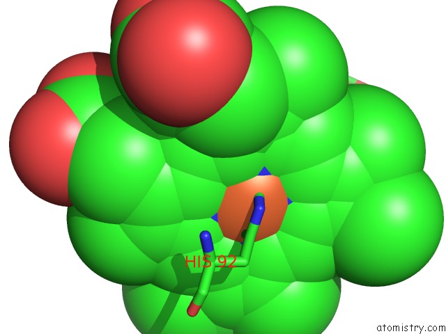
Mono view
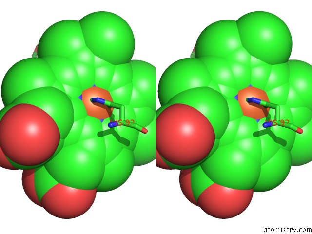
Stereo pair view
Mono view
Stereo pair view
A full contact list of Iron with other atoms in the Fe binding
site number 2 of Leghemoglobin A (Acetomet) within 5.0Å range:
|
Reference:
M.S.Hargrove,
J.K.Barry,
E.A.Brucker,
M.B.Berry,
G.N.Phillips Jr.,
J.S.Olson,
R.Arredondo-Peter,
J.M.Dean,
R.V.Klucas,
G.Sarath.
Characterization of Recombinant Soybean Leghemoglobin A and Apolar Distal Histidine Mutants. J.Mol.Biol. V. 266 1032 1997.
ISSN: ISSN 0022-2836
PubMed: 9086279
DOI: 10.1006/JMBI.1996.0833
Page generated: Sat Aug 3 02:48:34 2024
ISSN: ISSN 0022-2836
PubMed: 9086279
DOI: 10.1006/JMBI.1996.0833
Last articles
Cl in 5SDRCl in 5SDQ
Cl in 5SDP
Cl in 5SDN
Cl in 5SDO
Cl in 5SDM
Cl in 5SDL
Cl in 5SDK
Cl in 5SDI
Cl in 5SDJ