Iron »
PDB 1bj9-1c53 »
1bou »
Iron in PDB 1bou: Three-Dimensional Structure of Ligab
Enzymatic activity of Three-Dimensional Structure of Ligab
All present enzymatic activity of Three-Dimensional Structure of Ligab:
1.13.11.8;
1.13.11.8;
Protein crystallography data
The structure of Three-Dimensional Structure of Ligab, PDB code: 1bou
was solved by
K.Sugimoto,
T.Senda,
M.Fukuda,
Y.Mitsui,
with X-Ray Crystallography technique. A brief refinement statistics is given in the table below:
| Resolution Low / High (Å) | 60.00 / 2.20 |
| Space group | P 1 21 1 |
| Cell size a, b, c (Å), α, β, γ (°) | 65.400, 66.500, 119.800, 90.00, 92.50, 90.00 |
| R / Rfree (%) | 14.9 / 20.6 |
Iron Binding Sites:
The binding sites of Iron atom in the Three-Dimensional Structure of Ligab
(pdb code 1bou). This binding sites where shown within
5.0 Angstroms radius around Iron atom.
In total 2 binding sites of Iron where determined in the Three-Dimensional Structure of Ligab, PDB code: 1bou:
Jump to Iron binding site number: 1; 2;
In total 2 binding sites of Iron where determined in the Three-Dimensional Structure of Ligab, PDB code: 1bou:
Jump to Iron binding site number: 1; 2;
Iron binding site 1 out of 2 in 1bou
Go back to
Iron binding site 1 out
of 2 in the Three-Dimensional Structure of Ligab
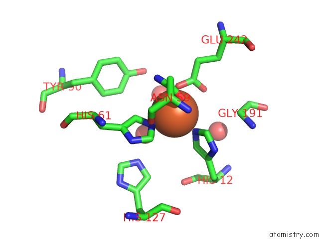
Mono view

Stereo pair view
Mono view
Stereo pair view
A full contact list of Iron with other atoms in the Fe binding
site number 1 of Three-Dimensional Structure of Ligab within 5.0Å range:
|
Iron binding site 2 out of 2 in 1bou
Go back to
Iron binding site 2 out
of 2 in the Three-Dimensional Structure of Ligab
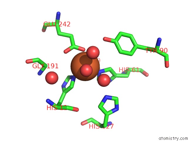
Mono view

Stereo pair view
Mono view
Stereo pair view
A full contact list of Iron with other atoms in the Fe binding
site number 2 of Three-Dimensional Structure of Ligab within 5.0Å range:
|
Reference:
K.Sugimoto,
T.Senda,
H.Aoshima,
E.Masai,
M.Fukuda,
Y.Mitsui.
Crystal Structure of An Aromatic Ring Opening Dioxygenase Ligab, A Protocatechuate 4,5-Dioxygenase, Under Aerobic Conditions. Structure V. 7 953 1999.
ISSN: ISSN 0969-2126
PubMed: 10467151
DOI: 10.1016/S0969-2126(99)80122-1
Page generated: Sat Aug 3 02:53:46 2024
ISSN: ISSN 0969-2126
PubMed: 10467151
DOI: 10.1016/S0969-2126(99)80122-1
Last articles
Zn in 9JYWZn in 9IR4
Zn in 9IR3
Zn in 9GMX
Zn in 9GMW
Zn in 9JEJ
Zn in 9ERF
Zn in 9ERE
Zn in 9EGV
Zn in 9EGW