Iron »
PDB 1bj9-1c53 »
1buw »
Iron in PDB 1buw: Crystal Structure of S-Nitroso-Nitrosyl Human Hemoglobin A
Protein crystallography data
The structure of Crystal Structure of S-Nitroso-Nitrosyl Human Hemoglobin A, PDB code: 1buw
was solved by
N.-L.Chan,
P.H.Rogers,
A.Arnone,
with X-Ray Crystallography technique. A brief refinement statistics is given in the table below:
| Resolution Low / High (Å) | 8.00 / 1.90 |
| Space group | P 21 21 21 |
| Cell size a, b, c (Å), α, β, γ (°) | 97.100, 101.200, 61.100, 90.00, 90.00, 90.00 |
| R / Rfree (%) | 18.4 / 24.4 |
Iron Binding Sites:
The binding sites of Iron atom in the Crystal Structure of S-Nitroso-Nitrosyl Human Hemoglobin A
(pdb code 1buw). This binding sites where shown within
5.0 Angstroms radius around Iron atom.
In total 4 binding sites of Iron where determined in the Crystal Structure of S-Nitroso-Nitrosyl Human Hemoglobin A, PDB code: 1buw:
Jump to Iron binding site number: 1; 2; 3; 4;
In total 4 binding sites of Iron where determined in the Crystal Structure of S-Nitroso-Nitrosyl Human Hemoglobin A, PDB code: 1buw:
Jump to Iron binding site number: 1; 2; 3; 4;
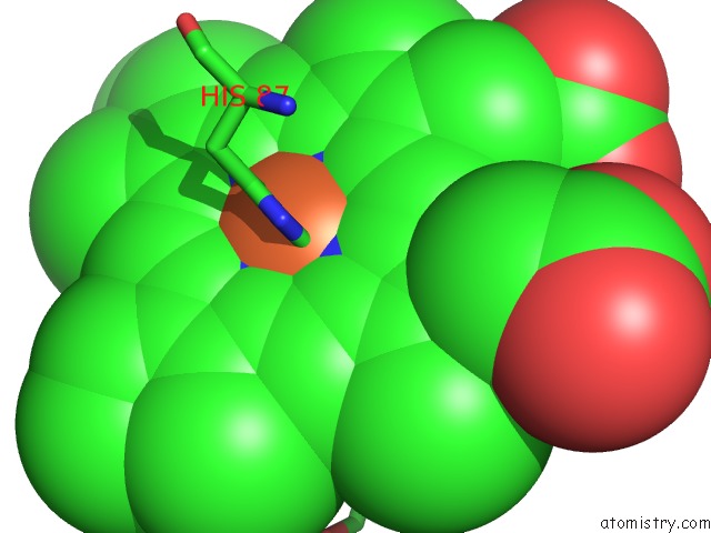
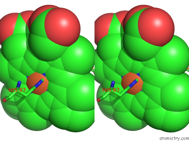
Iron binding site 1 out of 4 in 1buw
Go back to
Iron binding site 1 out
of 4 in the Crystal Structure of S-Nitroso-Nitrosyl Human Hemoglobin A
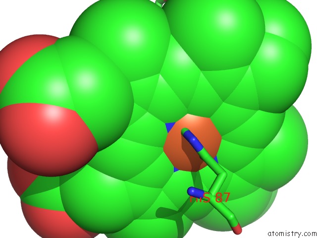
Mono view
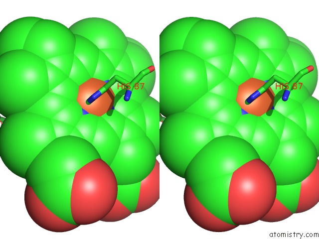
Stereo pair view
Mono view
Stereo pair view
A full contact list of Iron with other atoms in the Fe binding
site number 1 of Crystal Structure of S-Nitroso-Nitrosyl Human Hemoglobin A within 5.0Å range:
|
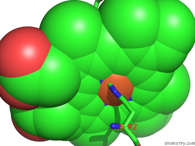
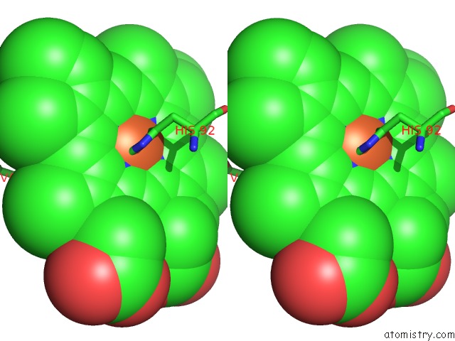
Iron binding site 2 out of 4 in 1buw
Go back to
Iron binding site 2 out
of 4 in the Crystal Structure of S-Nitroso-Nitrosyl Human Hemoglobin A
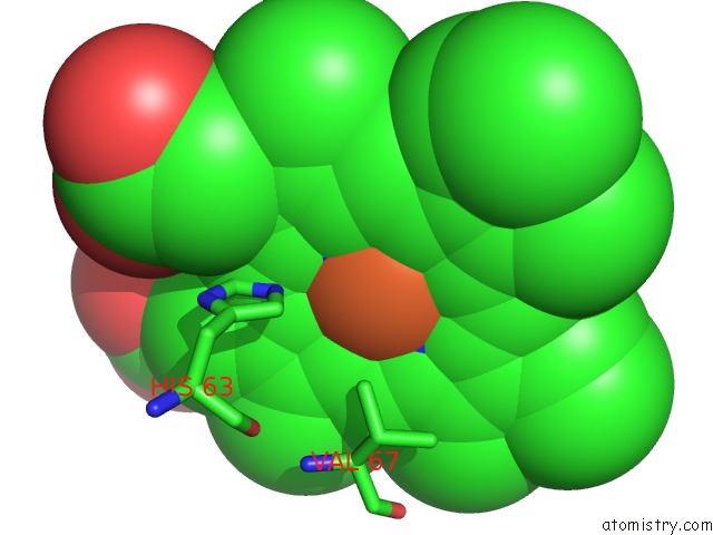
Mono view
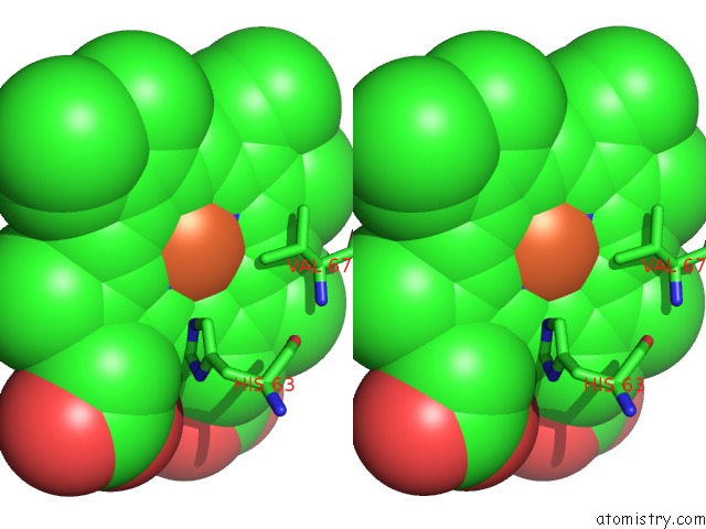
Stereo pair view
Mono view
Stereo pair view
A full contact list of Iron with other atoms in the Fe binding
site number 2 of Crystal Structure of S-Nitroso-Nitrosyl Human Hemoglobin A within 5.0Å range:
|
Iron binding site 3 out of 4 in 1buw
Go back to
Iron binding site 3 out
of 4 in the Crystal Structure of S-Nitroso-Nitrosyl Human Hemoglobin A
Mono view
Stereo pair view
Mono view
Stereo pair view
A full contact list of Iron with other atoms in the Fe binding
site number 3 of Crystal Structure of S-Nitroso-Nitrosyl Human Hemoglobin A within 5.0Å range:
|
Iron binding site 4 out of 4 in 1buw
Go back to
Iron binding site 4 out
of 4 in the Crystal Structure of S-Nitroso-Nitrosyl Human Hemoglobin A
Mono view
Stereo pair view
Mono view
Stereo pair view
A full contact list of Iron with other atoms in the Fe binding
site number 4 of Crystal Structure of S-Nitroso-Nitrosyl Human Hemoglobin A within 5.0Å range:
|
Reference:
N.L.Chan,
P.H.Rogers,
A.Arnone.
Crystal Structure of the S-Nitroso Form of Liganded Human Hemoglobin. Biochemistry V. 37 16459 1998.
ISSN: ISSN 0006-2960
PubMed: 9843411
DOI: 10.1021/BI9816711
Page generated: Sat Aug 3 02:55:44 2024
ISSN: ISSN 0006-2960
PubMed: 9843411
DOI: 10.1021/BI9816711
Last articles
Zn in 9J0NZn in 9J0O
Zn in 9J0P
Zn in 9FJX
Zn in 9EKB
Zn in 9C0F
Zn in 9CAH
Zn in 9CH0
Zn in 9CH3
Zn in 9CH1