Iron »
PDB 1bj9-1c53 »
1by5 »
Iron in PDB 1by5: Fhua From E. Coli, with Its Ligand Ferrichrome
Protein crystallography data
The structure of Fhua From E. Coli, with Its Ligand Ferrichrome, PDB code: 1by5
was solved by
K.P.Locher,
B.Rees,
R.Koebnik,
A.Mitschler,
L.Moulinier,
J.P.Rosenbusch,
D.Moras,
with X-Ray Crystallography technique. A brief refinement statistics is given in the table below:
| Resolution Low / High (Å) | 12.00 / 2.60 |
| Space group | C 1 2 1 |
| Cell size a, b, c (Å), α, β, γ (°) | 132.200, 89.400, 89.900, 90.00, 95.40, 90.00 |
| R / Rfree (%) | 18.4 / 22.9 |
Iron Binding Sites:
The binding sites of Iron atom in the Fhua From E. Coli, with Its Ligand Ferrichrome
(pdb code 1by5). This binding sites where shown within
5.0 Angstroms radius around Iron atom.
In total only one binding site of Iron was determined in the Fhua From E. Coli, with Its Ligand Ferrichrome, PDB code: 1by5:
In total only one binding site of Iron was determined in the Fhua From E. Coli, with Its Ligand Ferrichrome, PDB code: 1by5:
Iron binding site 1 out of 1 in 1by5
Go back to
Iron binding site 1 out
of 1 in the Fhua From E. Coli, with Its Ligand Ferrichrome
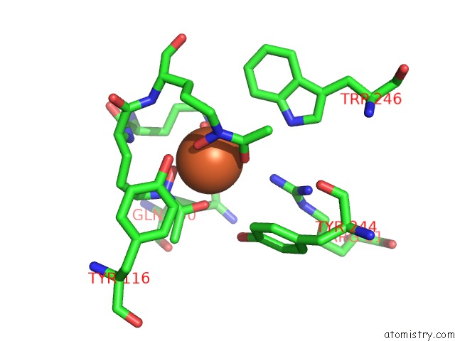
Mono view
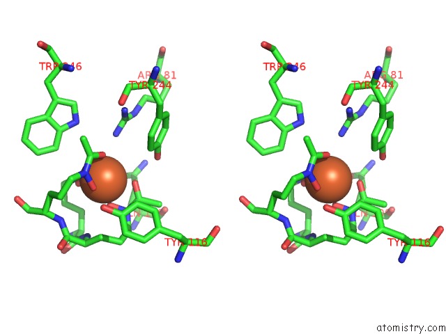
Stereo pair view
Mono view
Stereo pair view
A full contact list of Iron with other atoms in the Fe binding
site number 1 of Fhua From E. Coli, with Its Ligand Ferrichrome within 5.0Å range:
|
Reference:
K.P.Locher,
B.Rees,
R.Koebnik,
A.Mitschler,
L.Moulinier,
J.P.Rosenbusch,
D.Moras.
Transmembrane Signaling Across the Ligand-Gated Fhua Receptor: Crystal Structures of Free and Ferrichrome-Bound States Reveal Allosteric Changes. Cell(Cambridge,Mass.) V. 95 771 1998.
ISSN: ISSN 0092-8674
PubMed: 9865695
DOI: 10.1016/S0092-8674(00)81700-6
Page generated: Sat Aug 3 02:57:41 2024
ISSN: ISSN 0092-8674
PubMed: 9865695
DOI: 10.1016/S0092-8674(00)81700-6
Last articles
Zn in 9JYWZn in 9IR4
Zn in 9IR3
Zn in 9GMX
Zn in 9GMW
Zn in 9JEJ
Zn in 9ERF
Zn in 9ERE
Zn in 9EGV
Zn in 9EGW