Iron »
PDB 1c6o-1ch2 »
1cbl »
Iron in PDB 1cbl: The 1.9 Angstrom Structure of Deoxy-BETA4 Hemoglobin: Analysis of the Partitioning of Quaternary-Associated and Ligand-Induced Changes in Tertiary Structure
Protein crystallography data
The structure of The 1.9 Angstrom Structure of Deoxy-BETA4 Hemoglobin: Analysis of the Partitioning of Quaternary-Associated and Ligand-Induced Changes in Tertiary Structure, PDB code: 1cbl
was solved by
G.E.O.Borgstahl,
A.Arnone,
with X-Ray Crystallography technique. A brief refinement statistics is given in the table below:
| Resolution Low / High (Å) | 10.00 / 1.80 |
| Space group | P 1 21 1 |
| Cell size a, b, c (Å), α, β, γ (°) | 63.000, 81.800, 54.400, 90.00, 89.70, 90.00 |
| R / Rfree (%) | n/a / n/a |
Iron Binding Sites:
The binding sites of Iron atom in the The 1.9 Angstrom Structure of Deoxy-BETA4 Hemoglobin: Analysis of the Partitioning of Quaternary-Associated and Ligand-Induced Changes in Tertiary Structure
(pdb code 1cbl). This binding sites where shown within
5.0 Angstroms radius around Iron atom.
In total 4 binding sites of Iron where determined in the The 1.9 Angstrom Structure of Deoxy-BETA4 Hemoglobin: Analysis of the Partitioning of Quaternary-Associated and Ligand-Induced Changes in Tertiary Structure, PDB code: 1cbl:
Jump to Iron binding site number: 1; 2; 3; 4;
In total 4 binding sites of Iron where determined in the The 1.9 Angstrom Structure of Deoxy-BETA4 Hemoglobin: Analysis of the Partitioning of Quaternary-Associated and Ligand-Induced Changes in Tertiary Structure, PDB code: 1cbl:
Jump to Iron binding site number: 1; 2; 3; 4;
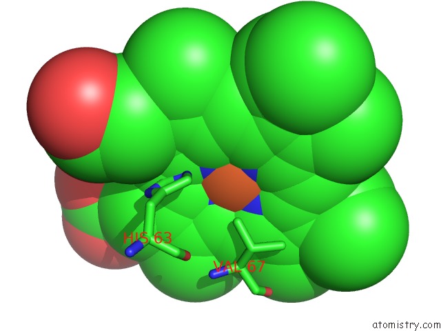
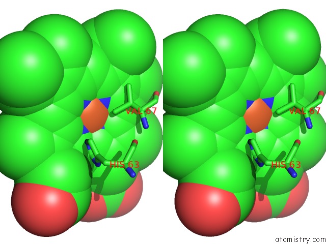
Iron binding site 1 out of 4 in 1cbl
Go back to
Iron binding site 1 out
of 4 in the The 1.9 Angstrom Structure of Deoxy-BETA4 Hemoglobin: Analysis of the Partitioning of Quaternary-Associated and Ligand-Induced Changes in Tertiary Structure
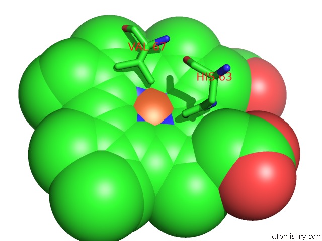
Mono view
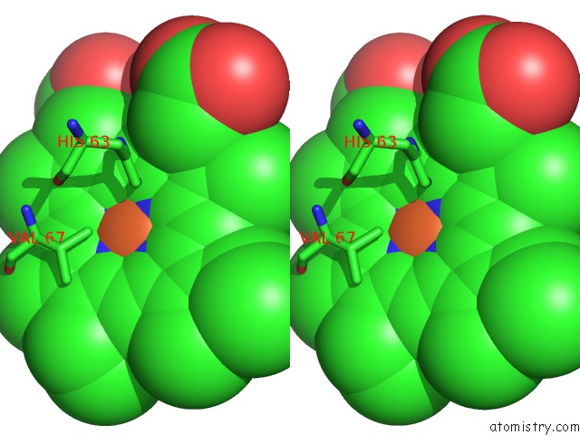
Stereo pair view
Mono view
Stereo pair view
A full contact list of Iron with other atoms in the Fe binding
site number 1 of The 1.9 Angstrom Structure of Deoxy-BETA4 Hemoglobin: Analysis of the Partitioning of Quaternary-Associated and Ligand-Induced Changes in Tertiary Structure within 5.0Å range:
|
Iron binding site 2 out of 4 in 1cbl
Go back to
Iron binding site 2 out
of 4 in the The 1.9 Angstrom Structure of Deoxy-BETA4 Hemoglobin: Analysis of the Partitioning of Quaternary-Associated and Ligand-Induced Changes in Tertiary Structure
Mono view
Stereo pair view
Mono view
Stereo pair view
A full contact list of Iron with other atoms in the Fe binding
site number 2 of The 1.9 Angstrom Structure of Deoxy-BETA4 Hemoglobin: Analysis of the Partitioning of Quaternary-Associated and Ligand-Induced Changes in Tertiary Structure within 5.0Å range:
|
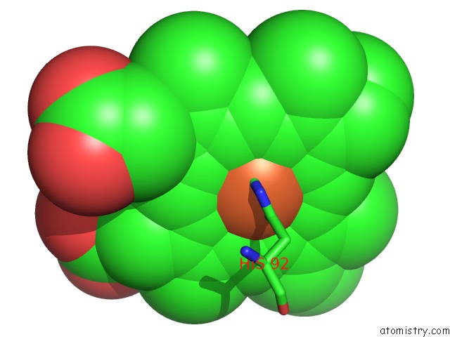
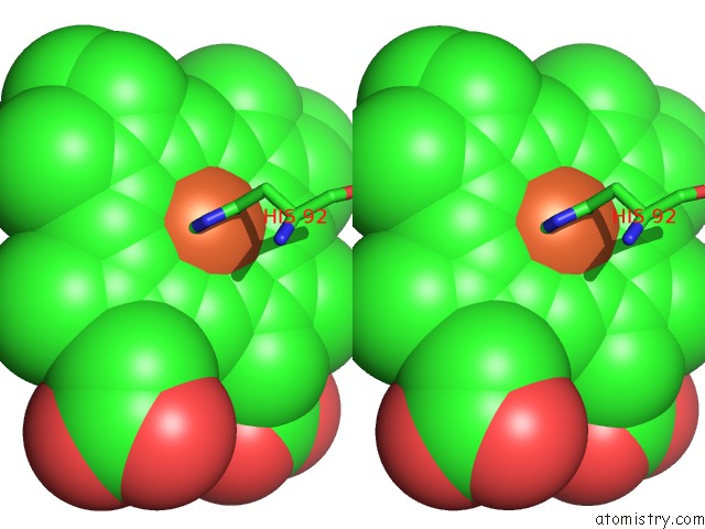
Iron binding site 3 out of 4 in 1cbl
Go back to
Iron binding site 3 out
of 4 in the The 1.9 Angstrom Structure of Deoxy-BETA4 Hemoglobin: Analysis of the Partitioning of Quaternary-Associated and Ligand-Induced Changes in Tertiary Structure
Mono view
Stereo pair view
Mono view
Stereo pair view
A full contact list of Iron with other atoms in the Fe binding
site number 3 of The 1.9 Angstrom Structure of Deoxy-BETA4 Hemoglobin: Analysis of the Partitioning of Quaternary-Associated and Ligand-Induced Changes in Tertiary Structure within 5.0Å range:
|
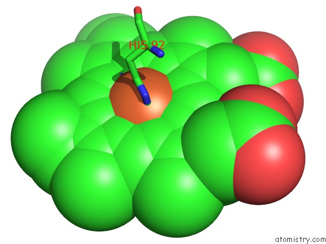
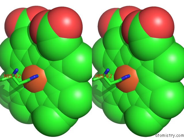
Iron binding site 4 out of 4 in 1cbl
Go back to
Iron binding site 4 out
of 4 in the The 1.9 Angstrom Structure of Deoxy-BETA4 Hemoglobin: Analysis of the Partitioning of Quaternary-Associated and Ligand-Induced Changes in Tertiary Structure
Mono view
Stereo pair view
Mono view
Stereo pair view
A full contact list of Iron with other atoms in the Fe binding
site number 4 of The 1.9 Angstrom Structure of Deoxy-BETA4 Hemoglobin: Analysis of the Partitioning of Quaternary-Associated and Ligand-Induced Changes in Tertiary Structure within 5.0Å range:
|
Reference:
G.E.Borgstahl,
P.H.Rogers,
A.Arnone.
The 1.9 A Structure of Deoxy Beta 4 Hemoglobin. Analysis of the Partitioning of Quaternary-Associated and Ligand-Induced Changes in Tertiary Structure. J.Mol.Biol. V. 236 831 1994.
ISSN: ISSN 0022-2836
PubMed: 8114097
DOI: 10.1006/JMBI.1994.1192
Page generated: Sat Aug 3 03:12:17 2024
ISSN: ISSN 0022-2836
PubMed: 8114097
DOI: 10.1006/JMBI.1994.1192
Last articles
Zn in 9JYWZn in 9IR4
Zn in 9IR3
Zn in 9GMX
Zn in 9GMW
Zn in 9JEJ
Zn in 9ERF
Zn in 9ERE
Zn in 9EGV
Zn in 9EGW