Iron »
PDB 1c6o-1ch2 »
1cbm »
Iron in PDB 1cbm: The 1.8 Angstrom Structure of Carbonmonoxy-BETA4 Hemoglobin: Analysis of A Homotetramer with the R Quaternary Structure of Liganded ALPHA2BETA2 Hemoglobin
Protein crystallography data
The structure of The 1.8 Angstrom Structure of Carbonmonoxy-BETA4 Hemoglobin: Analysis of A Homotetramer with the R Quaternary Structure of Liganded ALPHA2BETA2 Hemoglobin, PDB code: 1cbm
was solved by
G.E.O.Borgstahl,
A.Arnone,
with X-Ray Crystallography technique. A brief refinement statistics is given in the table below:
| Resolution Low / High (Å) | 10.00 / 1.74 |
| Space group | P 1 21 1 |
| Cell size a, b, c (Å), α, β, γ (°) | 63.300, 82.400, 53.700, 90.00, 90.10, 90.00 |
| R / Rfree (%) | n/a / n/a |
Iron Binding Sites:
The binding sites of Iron atom in the The 1.8 Angstrom Structure of Carbonmonoxy-BETA4 Hemoglobin: Analysis of A Homotetramer with the R Quaternary Structure of Liganded ALPHA2BETA2 Hemoglobin
(pdb code 1cbm). This binding sites where shown within
5.0 Angstroms radius around Iron atom.
In total 4 binding sites of Iron where determined in the The 1.8 Angstrom Structure of Carbonmonoxy-BETA4 Hemoglobin: Analysis of A Homotetramer with the R Quaternary Structure of Liganded ALPHA2BETA2 Hemoglobin, PDB code: 1cbm:
Jump to Iron binding site number: 1; 2; 3; 4;
In total 4 binding sites of Iron where determined in the The 1.8 Angstrom Structure of Carbonmonoxy-BETA4 Hemoglobin: Analysis of A Homotetramer with the R Quaternary Structure of Liganded ALPHA2BETA2 Hemoglobin, PDB code: 1cbm:
Jump to Iron binding site number: 1; 2; 3; 4;
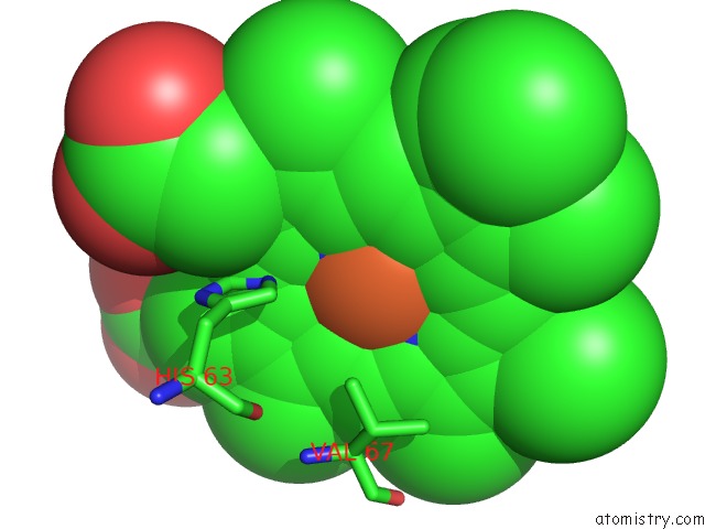
Iron binding site 1 out of 4 in 1cbm
Go back to
Iron binding site 1 out
of 4 in the The 1.8 Angstrom Structure of Carbonmonoxy-BETA4 Hemoglobin: Analysis of A Homotetramer with the R Quaternary Structure of Liganded ALPHA2BETA2 Hemoglobin
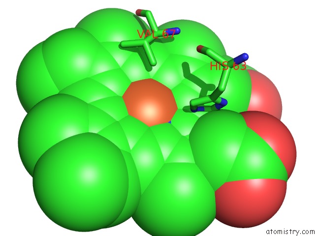
Mono view
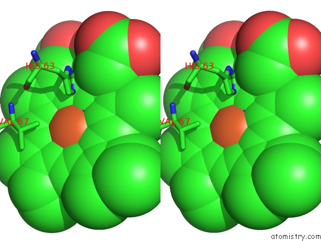
Stereo pair view
Mono view
Stereo pair view
A full contact list of Iron with other atoms in the Fe binding
site number 1 of The 1.8 Angstrom Structure of Carbonmonoxy-BETA4 Hemoglobin: Analysis of A Homotetramer with the R Quaternary Structure of Liganded ALPHA2BETA2 Hemoglobin within 5.0Å range:
|
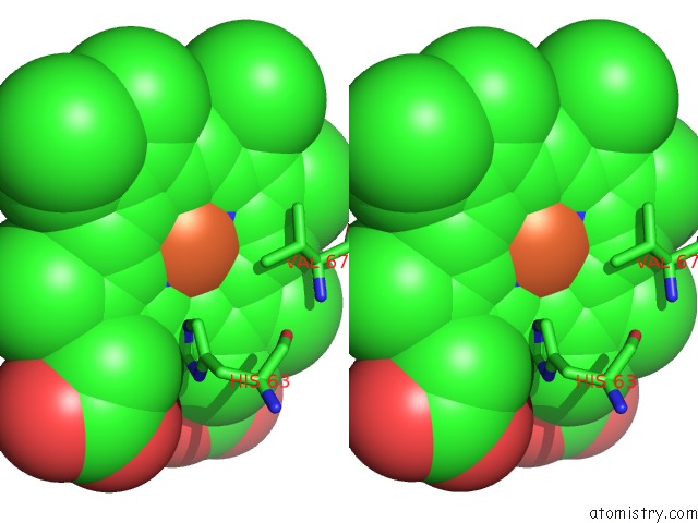
Iron binding site 2 out of 4 in 1cbm
Go back to
Iron binding site 2 out
of 4 in the The 1.8 Angstrom Structure of Carbonmonoxy-BETA4 Hemoglobin: Analysis of A Homotetramer with the R Quaternary Structure of Liganded ALPHA2BETA2 Hemoglobin
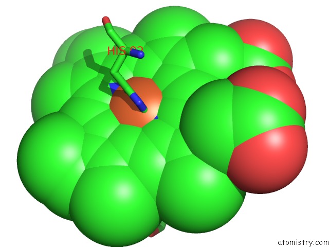
Mono view
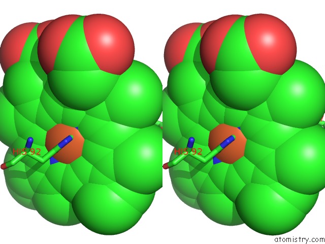
Stereo pair view
Mono view
Stereo pair view
A full contact list of Iron with other atoms in the Fe binding
site number 2 of The 1.8 Angstrom Structure of Carbonmonoxy-BETA4 Hemoglobin: Analysis of A Homotetramer with the R Quaternary Structure of Liganded ALPHA2BETA2 Hemoglobin within 5.0Å range:
|
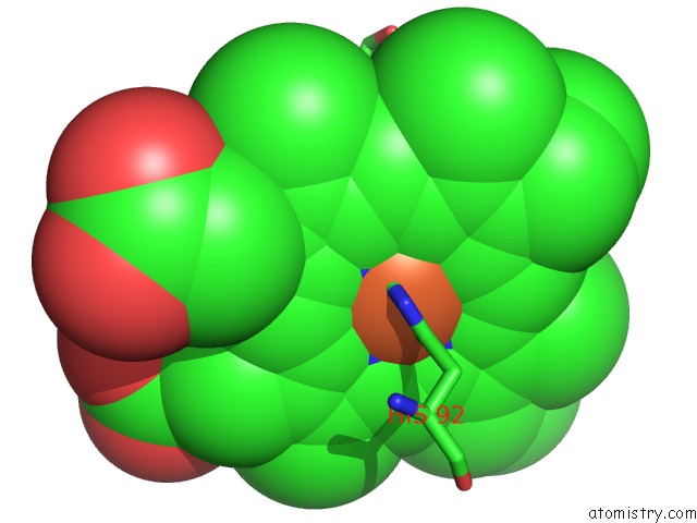
Iron binding site 3 out of 4 in 1cbm
Go back to
Iron binding site 3 out
of 4 in the The 1.8 Angstrom Structure of Carbonmonoxy-BETA4 Hemoglobin: Analysis of A Homotetramer with the R Quaternary Structure of Liganded ALPHA2BETA2 Hemoglobin
Mono view
Stereo pair view
Mono view
Stereo pair view
A full contact list of Iron with other atoms in the Fe binding
site number 3 of The 1.8 Angstrom Structure of Carbonmonoxy-BETA4 Hemoglobin: Analysis of A Homotetramer with the R Quaternary Structure of Liganded ALPHA2BETA2 Hemoglobin within 5.0Å range:
|
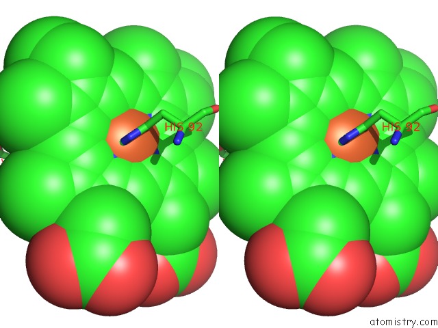
Iron binding site 4 out of 4 in 1cbm
Go back to
Iron binding site 4 out
of 4 in the The 1.8 Angstrom Structure of Carbonmonoxy-BETA4 Hemoglobin: Analysis of A Homotetramer with the R Quaternary Structure of Liganded ALPHA2BETA2 Hemoglobin
Mono view
Stereo pair view
Mono view
Stereo pair view
A full contact list of Iron with other atoms in the Fe binding
site number 4 of The 1.8 Angstrom Structure of Carbonmonoxy-BETA4 Hemoglobin: Analysis of A Homotetramer with the R Quaternary Structure of Liganded ALPHA2BETA2 Hemoglobin within 5.0Å range:
|
Reference:
G.E.Borgstahl,
P.H.Rogers,
A.Arnone.
The 1.8 A Structure of Carbonmonoxy-Beta 4 Hemoglobin. Analysis of A Homotetramer with the R Quaternary Structure of Liganded Alpha 2 Beta 2 Hemoglobin. J.Mol.Biol. V. 236 817 1994.
ISSN: ISSN 0022-2836
PubMed: 8114096
DOI: 10.1006/JMBI.1994.1191
Page generated: Sat Aug 3 03:12:44 2024
ISSN: ISSN 0022-2836
PubMed: 8114096
DOI: 10.1006/JMBI.1994.1191
Last articles
Zn in 9MJ5Zn in 9HNW
Zn in 9G0L
Zn in 9FNE
Zn in 9DZN
Zn in 9E0I
Zn in 9D32
Zn in 9DAK
Zn in 8ZXC
Zn in 8ZUF