Iron »
PDB 1c6o-1ch2 »
1cc1 »
Iron in PDB 1cc1: Crystal Structure of A Reduced, Active Form of the Ni-Fe-Se Hydrogenase From Desulfomicrobium Baculatum
Enzymatic activity of Crystal Structure of A Reduced, Active Form of the Ni-Fe-Se Hydrogenase From Desulfomicrobium Baculatum
All present enzymatic activity of Crystal Structure of A Reduced, Active Form of the Ni-Fe-Se Hydrogenase From Desulfomicrobium Baculatum:
1.18.99.1;
1.18.99.1;
Protein crystallography data
The structure of Crystal Structure of A Reduced, Active Form of the Ni-Fe-Se Hydrogenase From Desulfomicrobium Baculatum, PDB code: 1cc1
was solved by
E.Garcin,
X.Vernede,
E.C.Hatchikian,
A.Volbeda,
M.Frey,
J.C.Fontecilla-Camps,
with X-Ray Crystallography technique. A brief refinement statistics is given in the table below:
| Resolution Low / High (Å) | 8.00 / 2.15 |
| Space group | P 21 21 21 |
| Cell size a, b, c (Å), α, β, γ (°) | 110.390, 63.700, 99.580, 90.00, 90.00, 90.00 |
| R / Rfree (%) | 19.4 / 24.8 |
Other elements in 1cc1:
The structure of Crystal Structure of A Reduced, Active Form of the Ni-Fe-Se Hydrogenase From Desulfomicrobium Baculatum also contains other interesting chemical elements:
| Nickel | (Ni) | 1 atom |
Iron Binding Sites:
Pages:
>>> Page 1 <<< Page 2, Binding sites: 11 - 14;Binding sites:
The binding sites of Iron atom in the Crystal Structure of A Reduced, Active Form of the Ni-Fe-Se Hydrogenase From Desulfomicrobium Baculatum (pdb code 1cc1). This binding sites where shown within 5.0 Angstroms radius around Iron atom.In total 14 binding sites of Iron where determined in the Crystal Structure of A Reduced, Active Form of the Ni-Fe-Se Hydrogenase From Desulfomicrobium Baculatum, PDB code: 1cc1:
Jump to Iron binding site number: 1; 2; 3; 4; 5; 6; 7; 8; 9; 10;
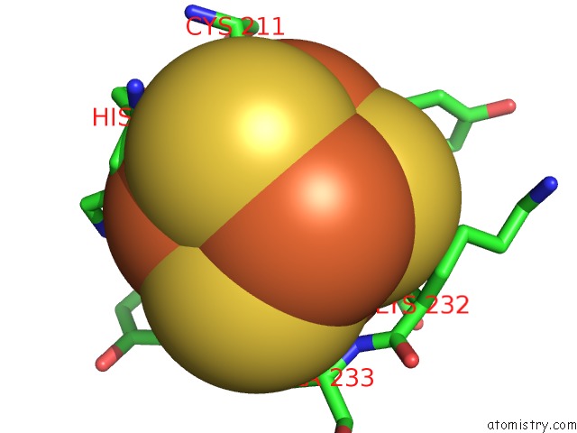
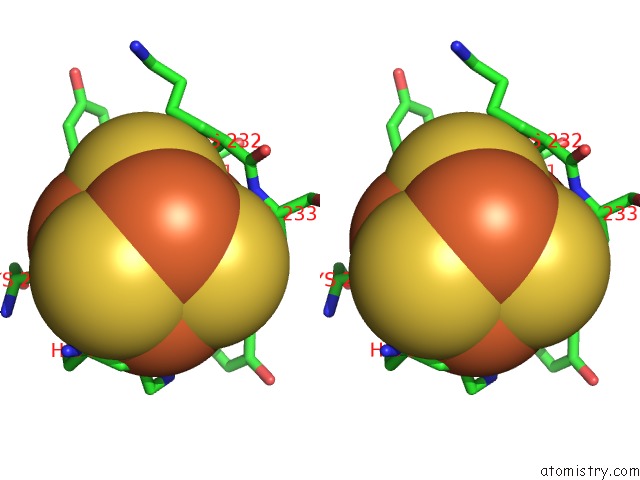
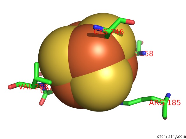
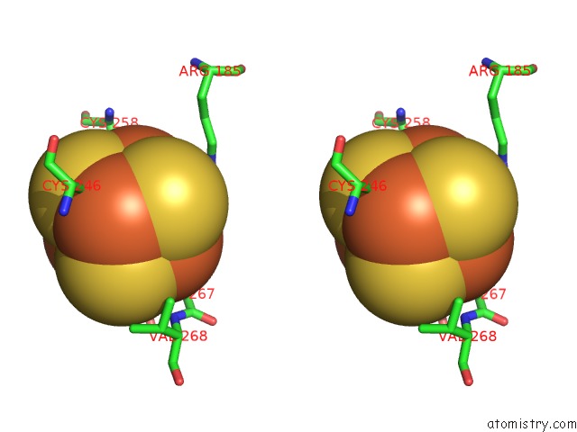
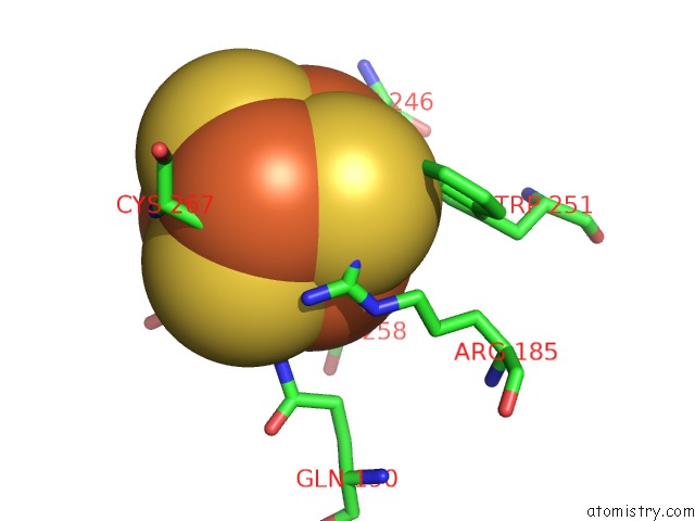
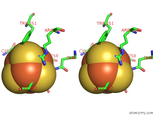
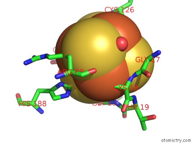
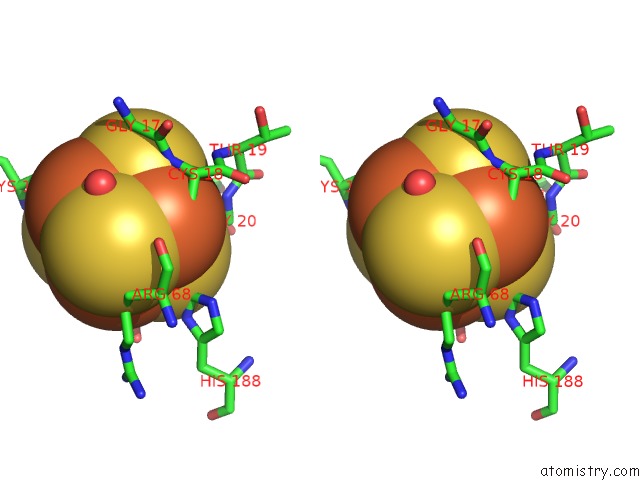
Iron binding site 1 out of 14 in 1cc1
Go back to
Iron binding site 1 out
of 14 in the Crystal Structure of A Reduced, Active Form of the Ni-Fe-Se Hydrogenase From Desulfomicrobium Baculatum
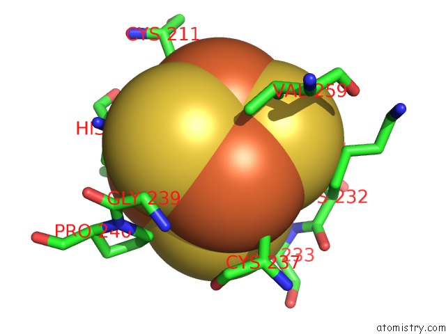
Mono view
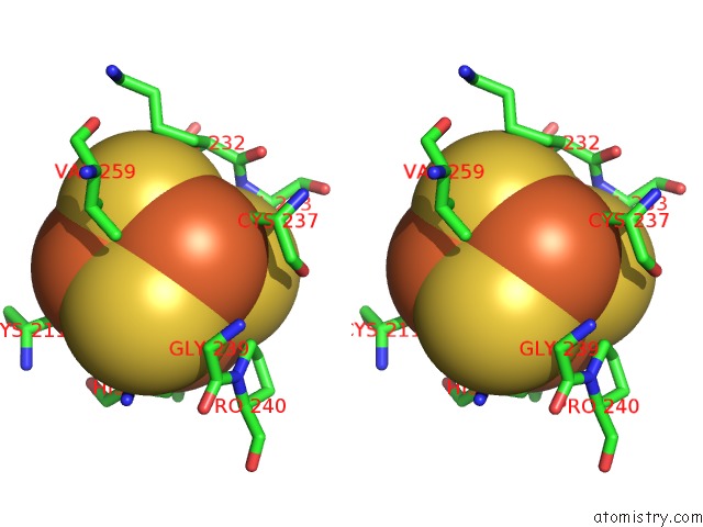
Stereo pair view
Mono view
Stereo pair view
A full contact list of Iron with other atoms in the Fe binding
site number 1 of Crystal Structure of A Reduced, Active Form of the Ni-Fe-Se Hydrogenase From Desulfomicrobium Baculatum within 5.0Å range:
|
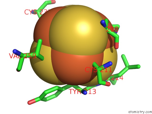
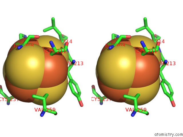
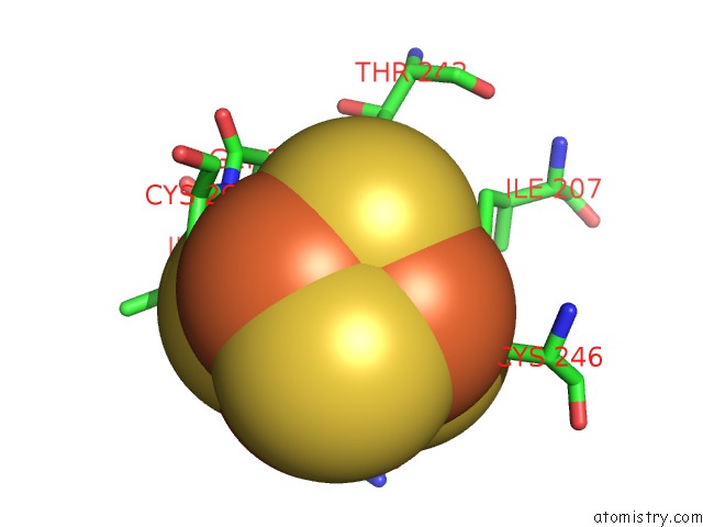
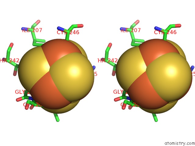
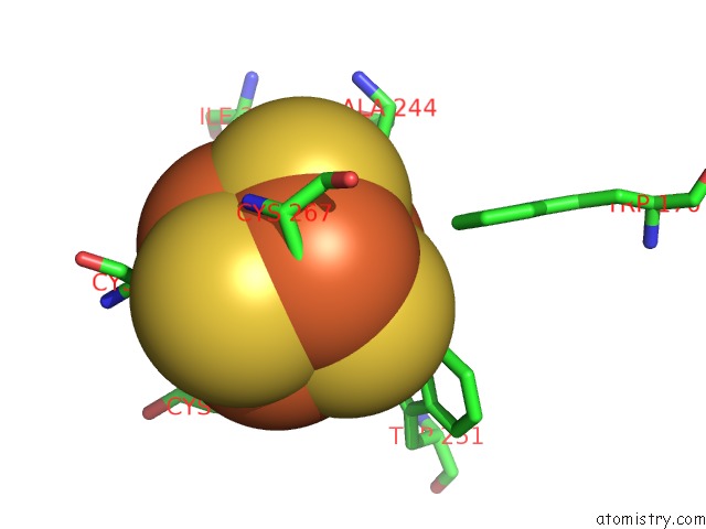
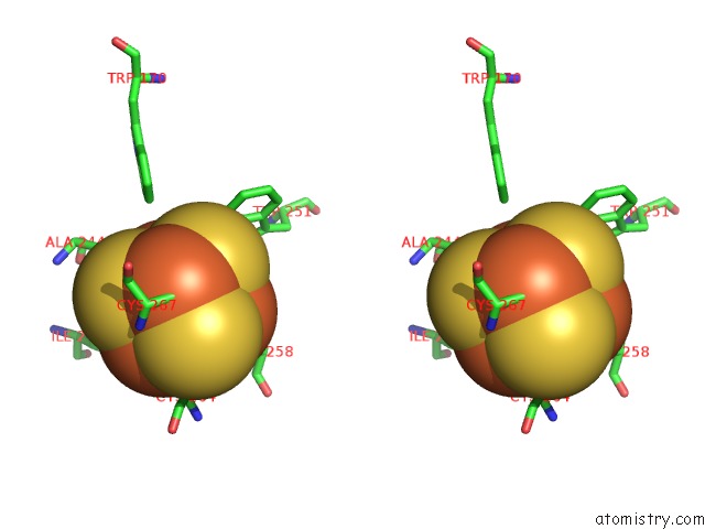
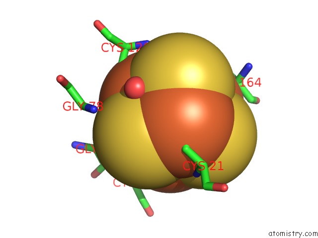
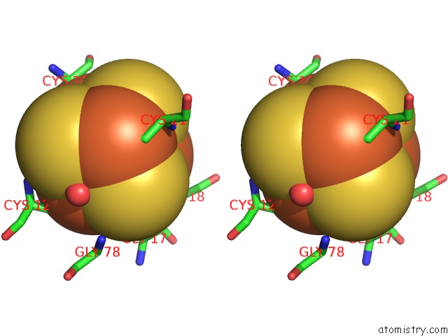
Iron binding site 2 out of 14 in 1cc1
Go back to
Iron binding site 2 out
of 14 in the Crystal Structure of A Reduced, Active Form of the Ni-Fe-Se Hydrogenase From Desulfomicrobium Baculatum
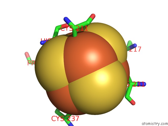
Mono view
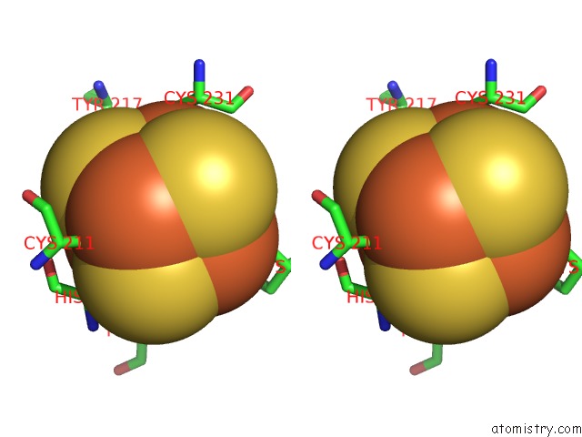
Stereo pair view
Mono view
Stereo pair view
A full contact list of Iron with other atoms in the Fe binding
site number 2 of Crystal Structure of A Reduced, Active Form of the Ni-Fe-Se Hydrogenase From Desulfomicrobium Baculatum within 5.0Å range:
|
Iron binding site 3 out of 14 in 1cc1
Go back to
Iron binding site 3 out
of 14 in the Crystal Structure of A Reduced, Active Form of the Ni-Fe-Se Hydrogenase From Desulfomicrobium Baculatum
Mono view
Stereo pair view
Mono view
Stereo pair view
A full contact list of Iron with other atoms in the Fe binding
site number 3 of Crystal Structure of A Reduced, Active Form of the Ni-Fe-Se Hydrogenase From Desulfomicrobium Baculatum within 5.0Å range:
|
Iron binding site 4 out of 14 in 1cc1
Go back to
Iron binding site 4 out
of 14 in the Crystal Structure of A Reduced, Active Form of the Ni-Fe-Se Hydrogenase From Desulfomicrobium Baculatum
Mono view
Stereo pair view
Mono view
Stereo pair view
A full contact list of Iron with other atoms in the Fe binding
site number 4 of Crystal Structure of A Reduced, Active Form of the Ni-Fe-Se Hydrogenase From Desulfomicrobium Baculatum within 5.0Å range:
|
Iron binding site 5 out of 14 in 1cc1
Go back to
Iron binding site 5 out
of 14 in the Crystal Structure of A Reduced, Active Form of the Ni-Fe-Se Hydrogenase From Desulfomicrobium Baculatum
Mono view
Stereo pair view
Mono view
Stereo pair view
A full contact list of Iron with other atoms in the Fe binding
site number 5 of Crystal Structure of A Reduced, Active Form of the Ni-Fe-Se Hydrogenase From Desulfomicrobium Baculatum within 5.0Å range:
|
Iron binding site 6 out of 14 in 1cc1
Go back to
Iron binding site 6 out
of 14 in the Crystal Structure of A Reduced, Active Form of the Ni-Fe-Se Hydrogenase From Desulfomicrobium Baculatum
Mono view
Stereo pair view
Mono view
Stereo pair view
A full contact list of Iron with other atoms in the Fe binding
site number 6 of Crystal Structure of A Reduced, Active Form of the Ni-Fe-Se Hydrogenase From Desulfomicrobium Baculatum within 5.0Å range:
|
Iron binding site 7 out of 14 in 1cc1
Go back to
Iron binding site 7 out
of 14 in the Crystal Structure of A Reduced, Active Form of the Ni-Fe-Se Hydrogenase From Desulfomicrobium Baculatum
Mono view
Stereo pair view
Mono view
Stereo pair view
A full contact list of Iron with other atoms in the Fe binding
site number 7 of Crystal Structure of A Reduced, Active Form of the Ni-Fe-Se Hydrogenase From Desulfomicrobium Baculatum within 5.0Å range:
|
Iron binding site 8 out of 14 in 1cc1
Go back to
Iron binding site 8 out
of 14 in the Crystal Structure of A Reduced, Active Form of the Ni-Fe-Se Hydrogenase From Desulfomicrobium Baculatum
Mono view
Stereo pair view
Mono view
Stereo pair view
A full contact list of Iron with other atoms in the Fe binding
site number 8 of Crystal Structure of A Reduced, Active Form of the Ni-Fe-Se Hydrogenase From Desulfomicrobium Baculatum within 5.0Å range:
|
Iron binding site 9 out of 14 in 1cc1
Go back to
Iron binding site 9 out
of 14 in the Crystal Structure of A Reduced, Active Form of the Ni-Fe-Se Hydrogenase From Desulfomicrobium Baculatum
Mono view
Stereo pair view
Mono view
Stereo pair view
A full contact list of Iron with other atoms in the Fe binding
site number 9 of Crystal Structure of A Reduced, Active Form of the Ni-Fe-Se Hydrogenase From Desulfomicrobium Baculatum within 5.0Å range:
|
Iron binding site 10 out of 14 in 1cc1
Go back to
Iron binding site 10 out
of 14 in the Crystal Structure of A Reduced, Active Form of the Ni-Fe-Se Hydrogenase From Desulfomicrobium Baculatum
Mono view
Stereo pair view
Mono view
Stereo pair view
A full contact list of Iron with other atoms in the Fe binding
site number 10 of Crystal Structure of A Reduced, Active Form of the Ni-Fe-Se Hydrogenase From Desulfomicrobium Baculatum within 5.0Å range:
|
Reference:
E.Garcin,
X.Vernede,
E.C.Hatchikian,
A.Volbeda,
M.Frey,
J.C.Fontecilla-Camps.
The Crystal Structure of A Reduced [Nifese] Hydrogenase Provides An Image of the Activated Catalytic Center Structure Fold.Des. V. 7 557 1999.
ISSN: ISSN 0969-2126
PubMed: 10378275
DOI: 10.1016/S0969-2126(99)80072-0
Page generated: Sat Aug 3 03:12:47 2024
ISSN: ISSN 0969-2126
PubMed: 10378275
DOI: 10.1016/S0969-2126(99)80072-0
Last articles
Zn in 9J0NZn in 9J0O
Zn in 9J0P
Zn in 9FJX
Zn in 9EKB
Zn in 9C0F
Zn in 9CAH
Zn in 9CH0
Zn in 9CH3
Zn in 9CH1