Iron »
PDB 1c6o-1ch2 »
1ce2 »
Iron in PDB 1ce2: Structure of Diferric Buffalo Lactoferrin at 2.5A Resolution
Protein crystallography data
The structure of Structure of Diferric Buffalo Lactoferrin at 2.5A Resolution, PDB code: 1ce2
was solved by
S.Karthikeyan,
M.Paramasivam,
S.Yadav,
A.Srinivasan,
T.P.Singh,
with X-Ray Crystallography technique. A brief refinement statistics is given in the table below:
| Resolution Low / High (Å) | 15.00 / 2.50 |
| Space group | P 21 21 21 |
| Cell size a, b, c (Å), α, β, γ (°) | 77.462, 91.035, 131.507, 90.00, 90.00, 90.00 |
| R / Rfree (%) | 18.7 / 26.5 |
Iron Binding Sites:
The binding sites of Iron atom in the Structure of Diferric Buffalo Lactoferrin at 2.5A Resolution
(pdb code 1ce2). This binding sites where shown within
5.0 Angstroms radius around Iron atom.
In total 2 binding sites of Iron where determined in the Structure of Diferric Buffalo Lactoferrin at 2.5A Resolution, PDB code: 1ce2:
Jump to Iron binding site number: 1; 2;
In total 2 binding sites of Iron where determined in the Structure of Diferric Buffalo Lactoferrin at 2.5A Resolution, PDB code: 1ce2:
Jump to Iron binding site number: 1; 2;
Iron binding site 1 out of 2 in 1ce2
Go back to
Iron binding site 1 out
of 2 in the Structure of Diferric Buffalo Lactoferrin at 2.5A Resolution
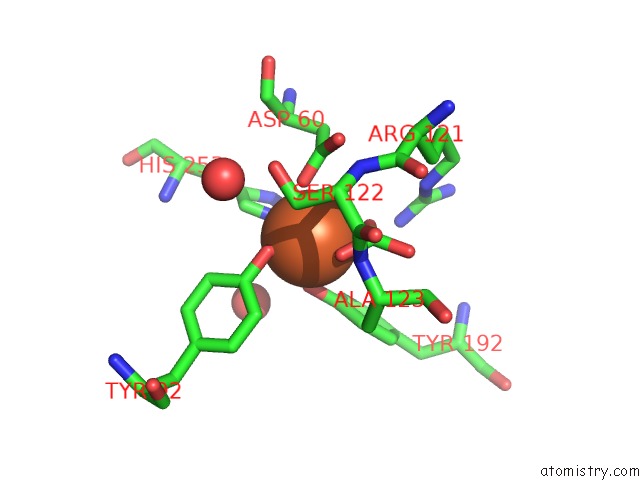
Mono view
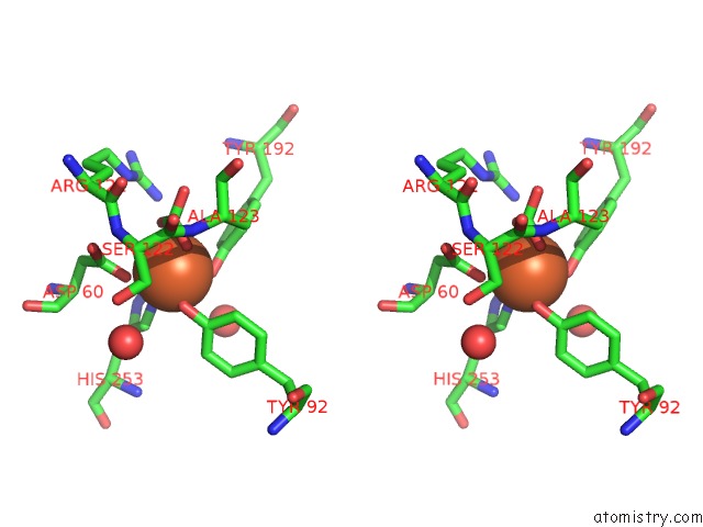
Stereo pair view
Mono view
Stereo pair view
A full contact list of Iron with other atoms in the Fe binding
site number 1 of Structure of Diferric Buffalo Lactoferrin at 2.5A Resolution within 5.0Å range:
|
Iron binding site 2 out of 2 in 1ce2
Go back to
Iron binding site 2 out
of 2 in the Structure of Diferric Buffalo Lactoferrin at 2.5A Resolution
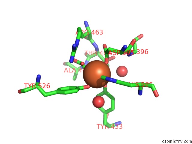
Mono view
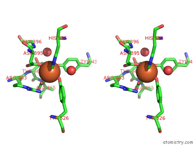
Stereo pair view
Mono view
Stereo pair view
A full contact list of Iron with other atoms in the Fe binding
site number 2 of Structure of Diferric Buffalo Lactoferrin at 2.5A Resolution within 5.0Å range:
|
Reference:
S.Karthikeyan,
M.Paramasivam,
S.Yadav,
A.Srinivasan,
T.P.Singh.
Structure of Buffalo Lactoferrin at 2.5 A Resolution Using Crystals Grown at 303 K Shows Different Orientations of the N and C Lobes. Acta Crystallogr.,Sect.D V. 55 1805 1999.
ISSN: ISSN 0907-4449
PubMed: 10531476
DOI: 10.1107/S0907444999010951
Page generated: Wed Jul 16 12:55:22 2025
ISSN: ISSN 0907-4449
PubMed: 10531476
DOI: 10.1107/S0907444999010951
Last articles
Fe in 2YXOFe in 2YRS
Fe in 2YXC
Fe in 2YNM
Fe in 2YVJ
Fe in 2YP1
Fe in 2YU2
Fe in 2YU1
Fe in 2YQB
Fe in 2YOO