Iron »
PDB 1c6o-1ch2 »
1cg8 »
Iron in PDB 1cg8: Co Form Hemoglobin From Dasyatis Akajei
Protein crystallography data
The structure of Co Form Hemoglobin From Dasyatis Akajei, PDB code: 1cg8
was solved by
K.T.Chong,
H.Morimoto,
with X-Ray Crystallography technique. A brief refinement statistics is given in the table below:
| Resolution Low / High (Å) | 6.00 / 1.90 |
| Space group | C 2 2 21 |
| Cell size a, b, c (Å), α, β, γ (°) | 57.660, 100.070, 107.610, 90.00, 90.00, 90.00 |
| R / Rfree (%) | 19.7 / 25.2 |
Iron Binding Sites:
The binding sites of Iron atom in the Co Form Hemoglobin From Dasyatis Akajei
(pdb code 1cg8). This binding sites where shown within
5.0 Angstroms radius around Iron atom.
In total 2 binding sites of Iron where determined in the Co Form Hemoglobin From Dasyatis Akajei, PDB code: 1cg8:
Jump to Iron binding site number: 1; 2;
In total 2 binding sites of Iron where determined in the Co Form Hemoglobin From Dasyatis Akajei, PDB code: 1cg8:
Jump to Iron binding site number: 1; 2;
Iron binding site 1 out of 2 in 1cg8
Go back to
Iron binding site 1 out
of 2 in the Co Form Hemoglobin From Dasyatis Akajei
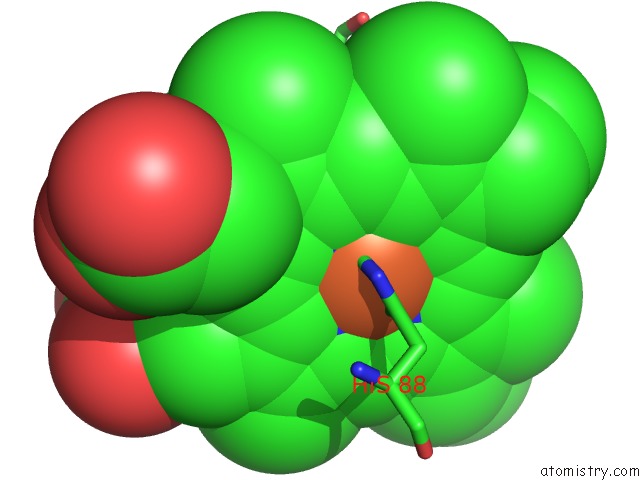
Mono view
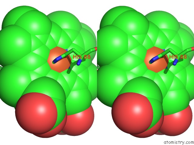
Stereo pair view
Mono view
Stereo pair view
A full contact list of Iron with other atoms in the Fe binding
site number 1 of Co Form Hemoglobin From Dasyatis Akajei within 5.0Å range:
|
Iron binding site 2 out of 2 in 1cg8
Go back to
Iron binding site 2 out
of 2 in the Co Form Hemoglobin From Dasyatis Akajei
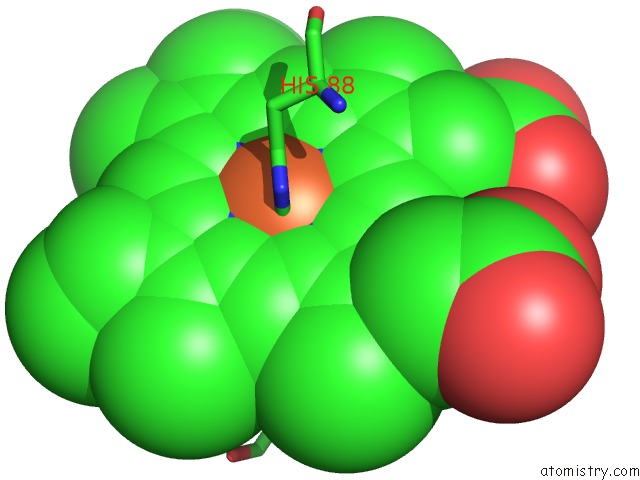
Mono view
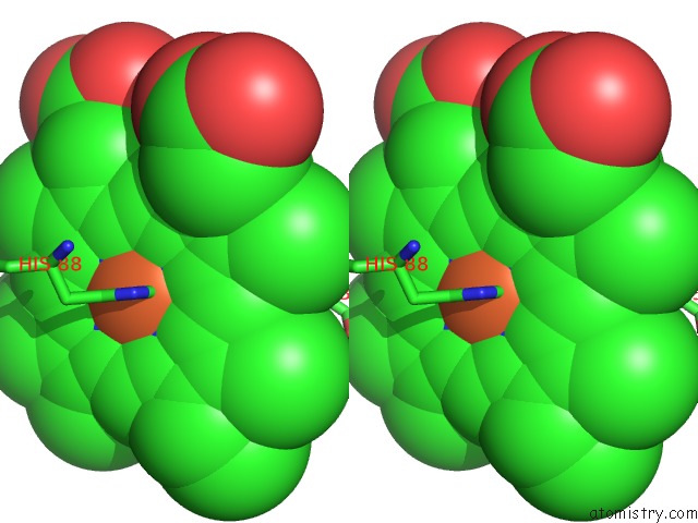
Stereo pair view
Mono view
Stereo pair view
A full contact list of Iron with other atoms in the Fe binding
site number 2 of Co Form Hemoglobin From Dasyatis Akajei within 5.0Å range:
|
Reference:
K.T.Chong,
G.Miyazaki,
H.Morimoto,
Y.Oda,
S.Y.Park.
Structures of the Deoxy and Co Forms of Haemoglobin From Dasyatis Akajei, A Cartilaginous Fish. Acta Crystallogr.,Sect.D V. 55 1291 1999.
ISSN: ISSN 0907-4449
PubMed: 10393295
DOI: 10.1107/S0907444999005934
Page generated: Sat Aug 3 03:16:39 2024
ISSN: ISSN 0907-4449
PubMed: 10393295
DOI: 10.1107/S0907444999005934
Last articles
Zn in 9J0NZn in 9J0O
Zn in 9J0P
Zn in 9FJX
Zn in 9EKB
Zn in 9C0F
Zn in 9CAH
Zn in 9CH0
Zn in 9CH3
Zn in 9CH1