Iron »
PDB 1d06-1dj5 »
1d3k »
Iron in PDB 1d3k: Human Serum Transferrin
Protein crystallography data
The structure of Human Serum Transferrin, PDB code: 1d3k
was solved by
H.-W.Yang,
R.T.A.Macgillivray,
J.Chen,
Y.Luo,
Y.Wang,
G.D.Brayer,
A.Mason,
R.C.Woodworth,
M.E.P.Murphy,
with X-Ray Crystallography technique. A brief refinement statistics is given in the table below:
| Resolution Low / High (Å) | 35.00 / 1.80 |
| Space group | P 21 21 21 |
| Cell size a, b, c (Å), α, β, γ (°) | 45.010, 57.810, 135.600, 90.00, 90.00, 90.00 |
| R / Rfree (%) | 18.4 / 22.5 |
Iron Binding Sites:
The binding sites of Iron atom in the Human Serum Transferrin
(pdb code 1d3k). This binding sites where shown within
5.0 Angstroms radius around Iron atom.
In total only one binding site of Iron was determined in the Human Serum Transferrin, PDB code: 1d3k:
In total only one binding site of Iron was determined in the Human Serum Transferrin, PDB code: 1d3k:
Iron binding site 1 out of 1 in 1d3k
Go back to
Iron binding site 1 out
of 1 in the Human Serum Transferrin
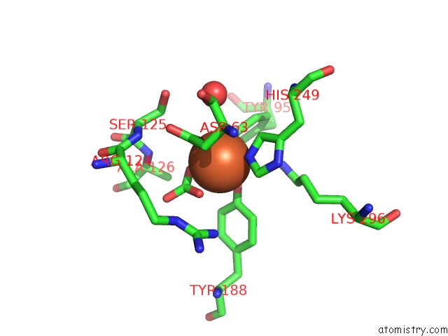
Mono view
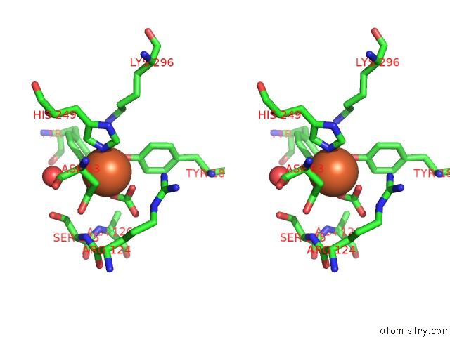
Stereo pair view
Mono view
Stereo pair view
A full contact list of Iron with other atoms in the Fe binding
site number 1 of Human Serum Transferrin within 5.0Å range:
|
Reference:
A.H.Yang,
R.T.Macgillivray,
J.Chen,
Y.Luo,
Y.Wang,
G.D.Brayer,
A.B.Mason,
R.C.Woodworth,
M.E.Murphy.
Crystal Structures of Two Mutants (K206Q, H207E) of the N-Lobe of Human Transferrin with Increased Affinity For Iron. Protein Sci. V. 9 49 2000.
ISSN: ISSN 0961-8368
PubMed: 10739246
Page generated: Sat Aug 3 03:35:47 2024
ISSN: ISSN 0961-8368
PubMed: 10739246
Last articles
Zn in 9J0NZn in 9J0O
Zn in 9J0P
Zn in 9FJX
Zn in 9EKB
Zn in 9C0F
Zn in 9CAH
Zn in 9CH0
Zn in 9CH3
Zn in 9CH1