Iron »
PDB 1d06-1dj5 »
1d3w »
Iron in PDB 1d3w: Crystal Structure of Ferredoxin 1 D15E Mutant From Azotobacter Vinelandii at 1.7 Angstrom Resolution.
Protein crystallography data
The structure of Crystal Structure of Ferredoxin 1 D15E Mutant From Azotobacter Vinelandii at 1.7 Angstrom Resolution., PDB code: 1d3w
was solved by
K.Chen,
J.Hirst,
R.Camba,
C.A.Bonagura,
C.D.Stout,
B.K.Burges,
F.A.Armstrong,
with X-Ray Crystallography technique. A brief refinement statistics is given in the table below:
| Resolution Low / High (Å) | 8.00 / 1.70 |
| Space group | P 41 21 2 |
| Cell size a, b, c (Å), α, β, γ (°) | 55.370, 55.370, 92.535, 90.00, 90.00, 90.00 |
| R / Rfree (%) | 20.9 / 24.7 |
Iron Binding Sites:
The binding sites of Iron atom in the Crystal Structure of Ferredoxin 1 D15E Mutant From Azotobacter Vinelandii at 1.7 Angstrom Resolution.
(pdb code 1d3w). This binding sites where shown within
5.0 Angstroms radius around Iron atom.
In total 7 binding sites of Iron where determined in the Crystal Structure of Ferredoxin 1 D15E Mutant From Azotobacter Vinelandii at 1.7 Angstrom Resolution., PDB code: 1d3w:
Jump to Iron binding site number: 1; 2; 3; 4; 5; 6; 7;
In total 7 binding sites of Iron where determined in the Crystal Structure of Ferredoxin 1 D15E Mutant From Azotobacter Vinelandii at 1.7 Angstrom Resolution., PDB code: 1d3w:
Jump to Iron binding site number: 1; 2; 3; 4; 5; 6; 7;
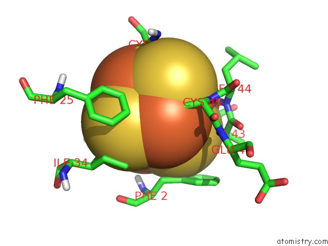
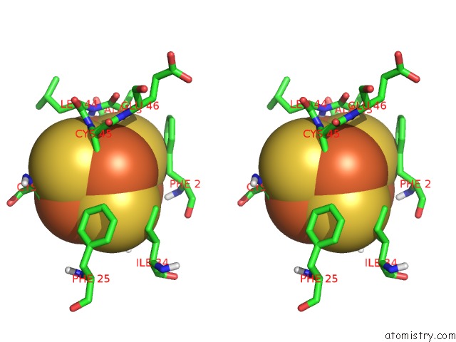
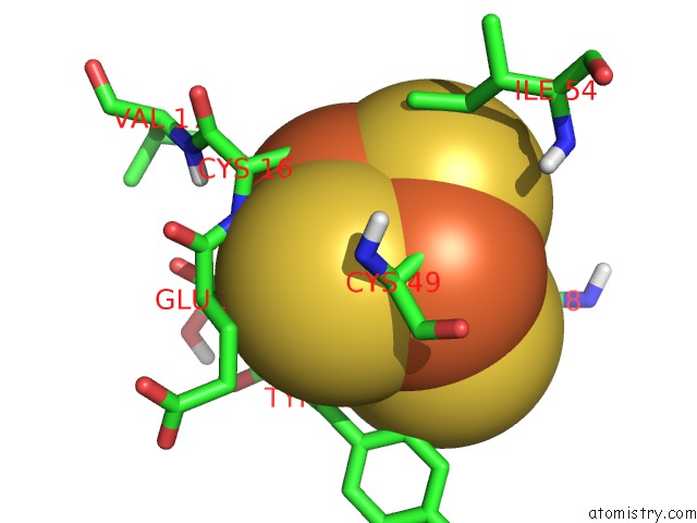
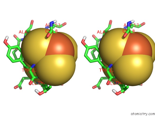
Iron binding site 1 out of 7 in 1d3w
Go back to
Iron binding site 1 out
of 7 in the Crystal Structure of Ferredoxin 1 D15E Mutant From Azotobacter Vinelandii at 1.7 Angstrom Resolution.
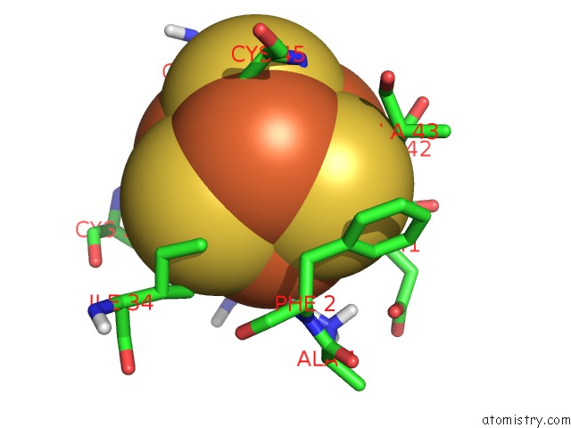
Mono view
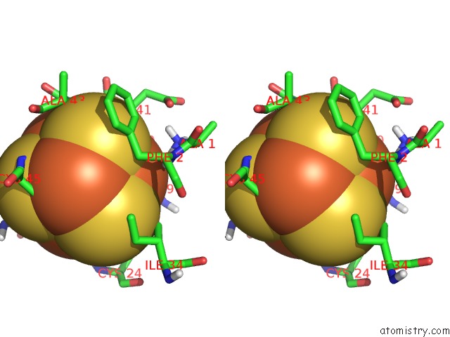
Stereo pair view
Mono view
Stereo pair view
A full contact list of Iron with other atoms in the Fe binding
site number 1 of Crystal Structure of Ferredoxin 1 D15E Mutant From Azotobacter Vinelandii at 1.7 Angstrom Resolution. within 5.0Å range:
|
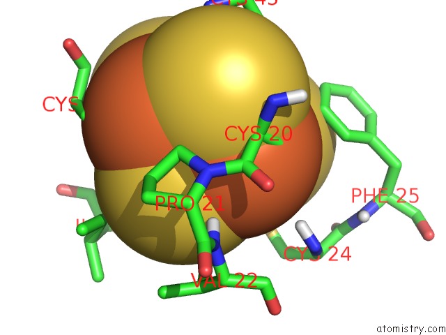
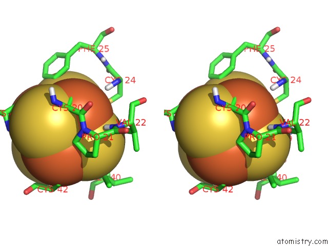
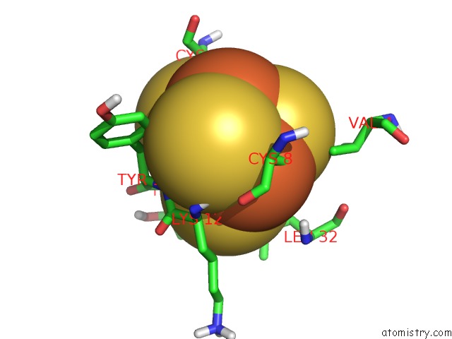
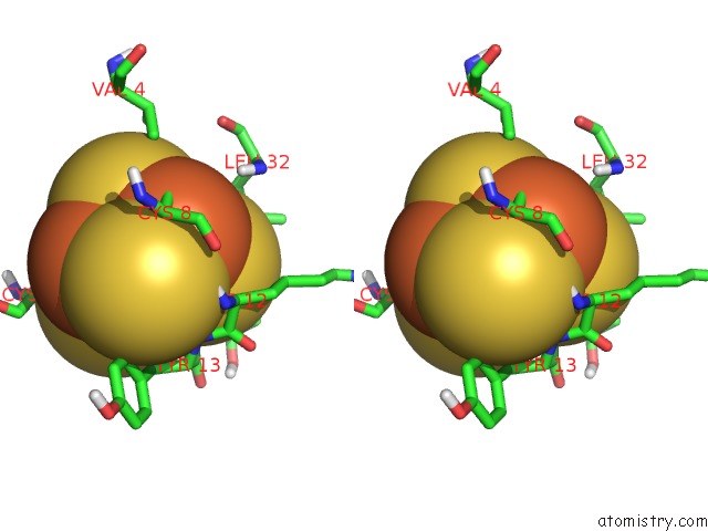
Iron binding site 2 out of 7 in 1d3w
Go back to
Iron binding site 2 out
of 7 in the Crystal Structure of Ferredoxin 1 D15E Mutant From Azotobacter Vinelandii at 1.7 Angstrom Resolution.
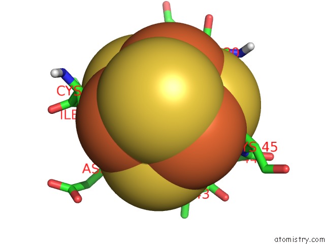
Mono view
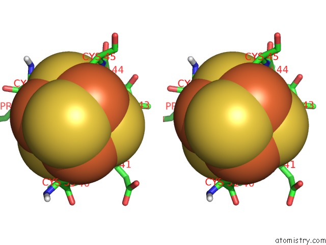
Stereo pair view
Mono view
Stereo pair view
A full contact list of Iron with other atoms in the Fe binding
site number 2 of Crystal Structure of Ferredoxin 1 D15E Mutant From Azotobacter Vinelandii at 1.7 Angstrom Resolution. within 5.0Å range:
|
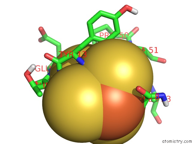
Iron binding site 3 out of 7 in 1d3w
Go back to
Iron binding site 3 out
of 7 in the Crystal Structure of Ferredoxin 1 D15E Mutant From Azotobacter Vinelandii at 1.7 Angstrom Resolution.
Mono view
Stereo pair view
Mono view
Stereo pair view
A full contact list of Iron with other atoms in the Fe binding
site number 3 of Crystal Structure of Ferredoxin 1 D15E Mutant From Azotobacter Vinelandii at 1.7 Angstrom Resolution. within 5.0Å range:
|
Iron binding site 4 out of 7 in 1d3w
Go back to
Iron binding site 4 out
of 7 in the Crystal Structure of Ferredoxin 1 D15E Mutant From Azotobacter Vinelandii at 1.7 Angstrom Resolution.
Mono view
Stereo pair view
Mono view
Stereo pair view
A full contact list of Iron with other atoms in the Fe binding
site number 4 of Crystal Structure of Ferredoxin 1 D15E Mutant From Azotobacter Vinelandii at 1.7 Angstrom Resolution. within 5.0Å range:
|
Iron binding site 5 out of 7 in 1d3w
Go back to
Iron binding site 5 out
of 7 in the Crystal Structure of Ferredoxin 1 D15E Mutant From Azotobacter Vinelandii at 1.7 Angstrom Resolution.
Mono view

Stereo pair view
Mono view
Stereo pair view
A full contact list of Iron with other atoms in the Fe binding
site number 5 of Crystal Structure of Ferredoxin 1 D15E Mutant From Azotobacter Vinelandii at 1.7 Angstrom Resolution. within 5.0Å range:
|
Iron binding site 6 out of 7 in 1d3w
Go back to
Iron binding site 6 out
of 7 in the Crystal Structure of Ferredoxin 1 D15E Mutant From Azotobacter Vinelandii at 1.7 Angstrom Resolution.
Mono view
Stereo pair view
Mono view
Stereo pair view
A full contact list of Iron with other atoms in the Fe binding
site number 6 of Crystal Structure of Ferredoxin 1 D15E Mutant From Azotobacter Vinelandii at 1.7 Angstrom Resolution. within 5.0Å range:
|
Iron binding site 7 out of 7 in 1d3w
Go back to
Iron binding site 7 out
of 7 in the Crystal Structure of Ferredoxin 1 D15E Mutant From Azotobacter Vinelandii at 1.7 Angstrom Resolution.
Mono view
Stereo pair view
Mono view
Stereo pair view
A full contact list of Iron with other atoms in the Fe binding
site number 7 of Crystal Structure of Ferredoxin 1 D15E Mutant From Azotobacter Vinelandii at 1.7 Angstrom Resolution. within 5.0Å range:
|
Reference:
K.Chen,
J.Hirst,
R.Camba,
C.A.Bonagura,
C.D.Stout,
B.K.Burgess,
F.A.Armstrong.
Atomically Defined Mechanism For Proton Transfer to A Buried Redox Centre in A Protein. Nature V. 405 814 2000.
ISSN: ISSN 0028-0836
PubMed: 10866206
DOI: 10.1038/35015610
Page generated: Sat Aug 3 03:36:55 2024
ISSN: ISSN 0028-0836
PubMed: 10866206
DOI: 10.1038/35015610
Last articles
F in 4OKXF in 4OKW
F in 4OKB
F in 4OKT
F in 4OK1
F in 4OJB
F in 4OJR
F in 4OI1
F in 4OJ9
F in 4OIU