Iron »
PDB 1d06-1dj5 »
1d5l »
Iron in PDB 1d5l: Crystal Structure of Cyanide-Bound Human Myeloperoxidase Isoform C at pH 5.5
Enzymatic activity of Crystal Structure of Cyanide-Bound Human Myeloperoxidase Isoform C at pH 5.5
All present enzymatic activity of Crystal Structure of Cyanide-Bound Human Myeloperoxidase Isoform C at pH 5.5:
1.11.1.7;
1.11.1.7;
Protein crystallography data
The structure of Crystal Structure of Cyanide-Bound Human Myeloperoxidase Isoform C at pH 5.5, PDB code: 1d5l
was solved by
T.J.Fiedler,
C.A.Davey,
R.E.Fenna,
with X-Ray Crystallography technique. A brief refinement statistics is given in the table below:
| Resolution Low / High (Å) | 30.00 / 1.90 |
| Space group | P 1 21 1 |
| Cell size a, b, c (Å), α, β, γ (°) | 111.215, 63.507, 92.337, 90.00, 97.43, 90.00 |
| R / Rfree (%) | 17.2 / 21.5 |
Other elements in 1d5l:
The structure of Crystal Structure of Cyanide-Bound Human Myeloperoxidase Isoform C at pH 5.5 also contains other interesting chemical elements:
| Chlorine | (Cl) | 2 atoms |
| Calcium | (Ca) | 2 atoms |
Iron Binding Sites:
The binding sites of Iron atom in the Crystal Structure of Cyanide-Bound Human Myeloperoxidase Isoform C at pH 5.5
(pdb code 1d5l). This binding sites where shown within
5.0 Angstroms radius around Iron atom.
In total 2 binding sites of Iron where determined in the Crystal Structure of Cyanide-Bound Human Myeloperoxidase Isoform C at pH 5.5, PDB code: 1d5l:
Jump to Iron binding site number: 1; 2;
In total 2 binding sites of Iron where determined in the Crystal Structure of Cyanide-Bound Human Myeloperoxidase Isoform C at pH 5.5, PDB code: 1d5l:
Jump to Iron binding site number: 1; 2;
Iron binding site 1 out of 2 in 1d5l
Go back to
Iron binding site 1 out
of 2 in the Crystal Structure of Cyanide-Bound Human Myeloperoxidase Isoform C at pH 5.5
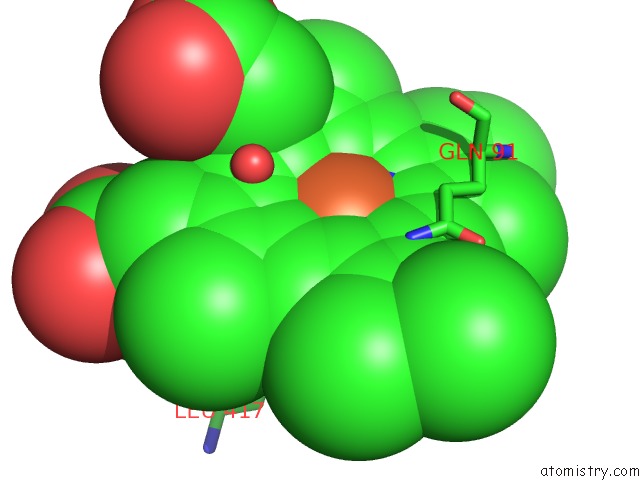
Mono view
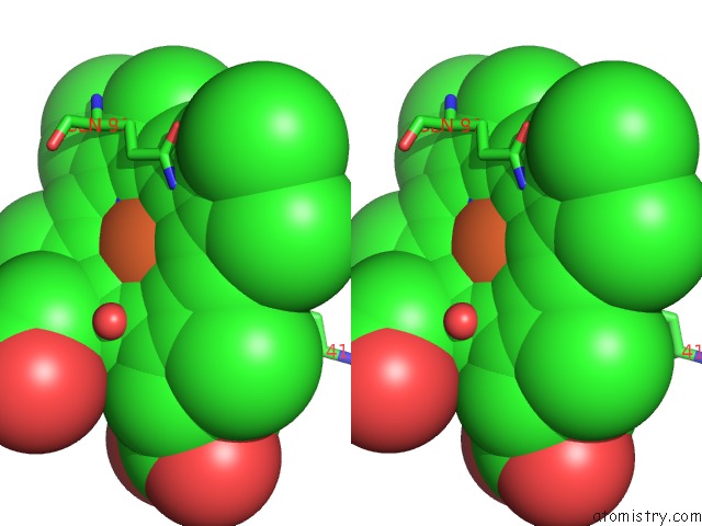
Stereo pair view
Mono view
Stereo pair view
A full contact list of Iron with other atoms in the Fe binding
site number 1 of Crystal Structure of Cyanide-Bound Human Myeloperoxidase Isoform C at pH 5.5 within 5.0Å range:
|
Iron binding site 2 out of 2 in 1d5l
Go back to
Iron binding site 2 out
of 2 in the Crystal Structure of Cyanide-Bound Human Myeloperoxidase Isoform C at pH 5.5
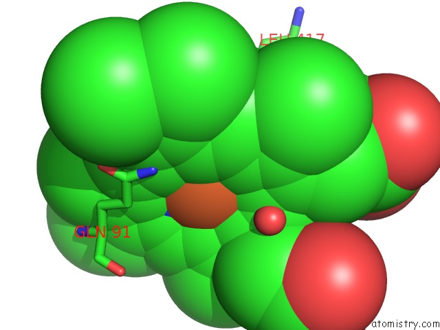
Mono view
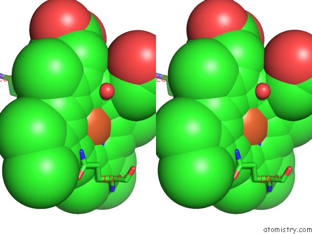
Stereo pair view
Mono view
Stereo pair view
A full contact list of Iron with other atoms in the Fe binding
site number 2 of Crystal Structure of Cyanide-Bound Human Myeloperoxidase Isoform C at pH 5.5 within 5.0Å range:
|
Reference:
M.Blair-Johnson,
T.Fiedler,
R.Fenna.
Human Myeloperoxidase: Structure of A Cyanide Complex and Its Interaction with Bromide and Thiocyanate Substrates at 1.9 A Resolution. Biochemistry V. 40 13990 2001.
ISSN: ISSN 0006-2960
PubMed: 11705390
DOI: 10.1021/BI0111808
Page generated: Wed Jul 16 13:11:45 2025
ISSN: ISSN 0006-2960
PubMed: 11705390
DOI: 10.1021/BI0111808
Last articles
Fe in 2YXOFe in 2YRS
Fe in 2YXC
Fe in 2YNM
Fe in 2YVJ
Fe in 2YP1
Fe in 2YU2
Fe in 2YU1
Fe in 2YQB
Fe in 2YOO