Iron »
PDB 1dj7-1dry »
1dj7 »
Iron in PDB 1dj7: Crystal Structure of Ferredoxin Thioredoxin Reductase
Protein crystallography data
The structure of Crystal Structure of Ferredoxin Thioredoxin Reductase, PDB code: 1dj7
was solved by
S.Dai,
C.Schwendtmayer,
P.Schurmann,
S.Ramaswamy,
H.Eklund,
with X-Ray Crystallography technique. A brief refinement statistics is given in the table below:
| Resolution Low / High (Å) | 10.00 / 1.60 |
| Space group | P 43 21 2 |
| Cell size a, b, c (Å), α, β, γ (°) | 45.296, 45.296, 172.147, 90.00, 90.00, 90.00 |
| R / Rfree (%) | 23.8 / 27.8 |
Iron Binding Sites:
The binding sites of Iron atom in the Crystal Structure of Ferredoxin Thioredoxin Reductase
(pdb code 1dj7). This binding sites where shown within
5.0 Angstroms radius around Iron atom.
In total 4 binding sites of Iron where determined in the Crystal Structure of Ferredoxin Thioredoxin Reductase, PDB code: 1dj7:
Jump to Iron binding site number: 1; 2; 3; 4;
In total 4 binding sites of Iron where determined in the Crystal Structure of Ferredoxin Thioredoxin Reductase, PDB code: 1dj7:
Jump to Iron binding site number: 1; 2; 3; 4;
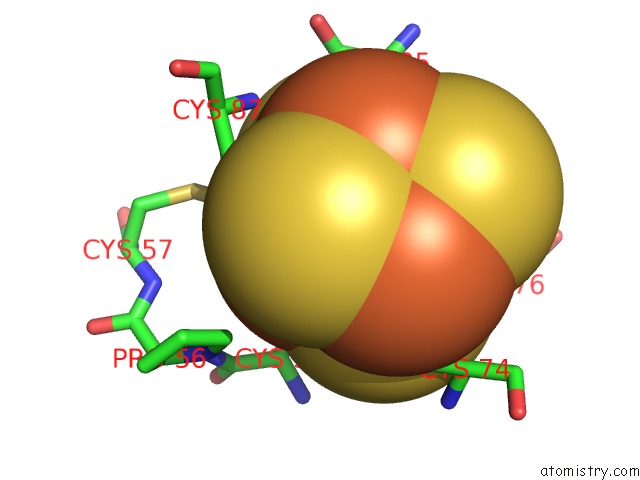
Iron binding site 1 out of 4 in 1dj7
Go back to
Iron binding site 1 out
of 4 in the Crystal Structure of Ferredoxin Thioredoxin Reductase
Mono view

Stereo pair view
Mono view
Stereo pair view
A full contact list of Iron with other atoms in the Fe binding
site number 1 of Crystal Structure of Ferredoxin Thioredoxin Reductase within 5.0Å range:
|
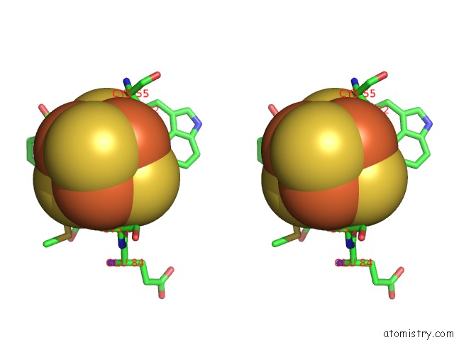
Iron binding site 2 out of 4 in 1dj7
Go back to
Iron binding site 2 out
of 4 in the Crystal Structure of Ferredoxin Thioredoxin Reductase

Mono view
Stereo pair view
Mono view
Stereo pair view
A full contact list of Iron with other atoms in the Fe binding
site number 2 of Crystal Structure of Ferredoxin Thioredoxin Reductase within 5.0Å range:
|
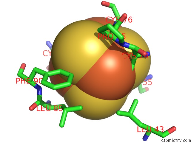
Iron binding site 3 out of 4 in 1dj7
Go back to
Iron binding site 3 out
of 4 in the Crystal Structure of Ferredoxin Thioredoxin Reductase

Mono view

Stereo pair view
Mono view
Stereo pair view
A full contact list of Iron with other atoms in the Fe binding
site number 3 of Crystal Structure of Ferredoxin Thioredoxin Reductase within 5.0Å range:
|
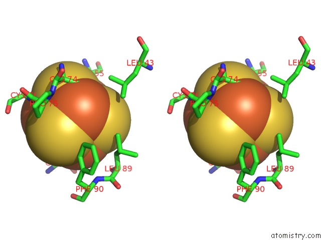
Iron binding site 4 out of 4 in 1dj7
Go back to
Iron binding site 4 out
of 4 in the Crystal Structure of Ferredoxin Thioredoxin Reductase
Mono view
Stereo pair view
Mono view
Stereo pair view
A full contact list of Iron with other atoms in the Fe binding
site number 4 of Crystal Structure of Ferredoxin Thioredoxin Reductase within 5.0Å range:
|
Reference:
S.Dai,
C.Schwendtmayer,
P.Schurmann,
S.Ramaswamy,
H.Eklund.
Redox Signaling in Chloroplasts: Cleavage of Disulfides By An Iron-Sulfur Cluster. Science V. 287 655 2000.
ISSN: ISSN 0036-8075
PubMed: 10649999
DOI: 10.1126/SCIENCE.287.5453.655
Page generated: Sat Aug 3 03:48:37 2024
ISSN: ISSN 0036-8075
PubMed: 10649999
DOI: 10.1126/SCIENCE.287.5453.655
Last articles
Zn in 9JYWZn in 9IR4
Zn in 9IR3
Zn in 9GMX
Zn in 9GMW
Zn in 9JEJ
Zn in 9ERF
Zn in 9ERE
Zn in 9EGV
Zn in 9EGW