Iron »
PDB 1dj7-1dry »
1dk0 »
Iron in PDB 1dk0: Crystal Structure of the Hemophore Hasa From Serratia Marcescens Crystal Form P2(1), PH8
Protein crystallography data
The structure of Crystal Structure of the Hemophore Hasa From Serratia Marcescens Crystal Form P2(1), PH8, PDB code: 1dk0
was solved by
P.Arnoux,
R.Haser,
N.Izadi-Pruneyre,
A.Lecroisey,
M.Czjzek,
with X-Ray Crystallography technique. A brief refinement statistics is given in the table below:
| Resolution Low / High (Å) | 15.00 / 1.77 |
| Space group | P 1 21 1 |
| Cell size a, b, c (Å), α, β, γ (°) | 45.540, 66.210, 58.930, 90.00, 104.91, 90.00 |
| R / Rfree (%) | 16.9 / 21.3 |
Iron Binding Sites:
The binding sites of Iron atom in the Crystal Structure of the Hemophore Hasa From Serratia Marcescens Crystal Form P2(1), PH8
(pdb code 1dk0). This binding sites where shown within
5.0 Angstroms radius around Iron atom.
In total 2 binding sites of Iron where determined in the Crystal Structure of the Hemophore Hasa From Serratia Marcescens Crystal Form P2(1), PH8, PDB code: 1dk0:
Jump to Iron binding site number: 1; 2;
In total 2 binding sites of Iron where determined in the Crystal Structure of the Hemophore Hasa From Serratia Marcescens Crystal Form P2(1), PH8, PDB code: 1dk0:
Jump to Iron binding site number: 1; 2;
Iron binding site 1 out of 2 in 1dk0
Go back to
Iron binding site 1 out
of 2 in the Crystal Structure of the Hemophore Hasa From Serratia Marcescens Crystal Form P2(1), PH8
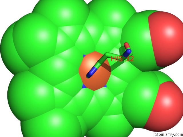
Mono view
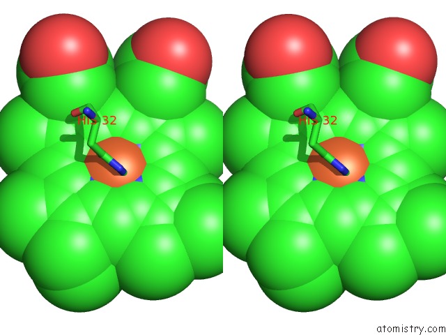
Stereo pair view
Mono view
Stereo pair view
A full contact list of Iron with other atoms in the Fe binding
site number 1 of Crystal Structure of the Hemophore Hasa From Serratia Marcescens Crystal Form P2(1), PH8 within 5.0Å range:
|
Iron binding site 2 out of 2 in 1dk0
Go back to
Iron binding site 2 out
of 2 in the Crystal Structure of the Hemophore Hasa From Serratia Marcescens Crystal Form P2(1), PH8
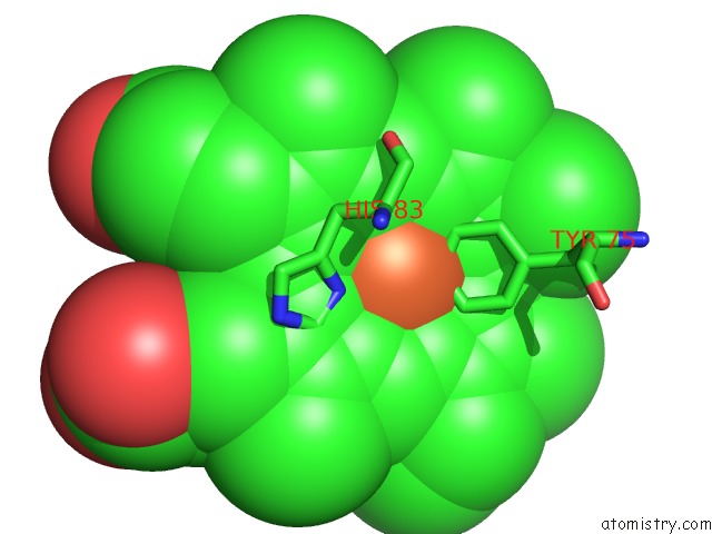
Mono view
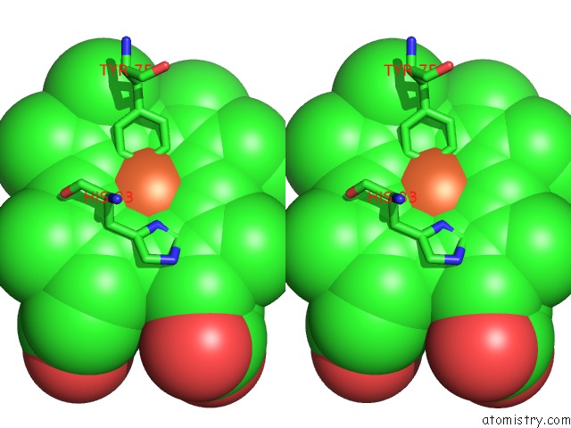
Stereo pair view
Mono view
Stereo pair view
A full contact list of Iron with other atoms in the Fe binding
site number 2 of Crystal Structure of the Hemophore Hasa From Serratia Marcescens Crystal Form P2(1), PH8 within 5.0Å range:
|
Reference:
P.Arnoux,
R.Haser,
N.Izadi-Pruneyre,
A.Lecroisey,
M.Czjzek.
Functional Aspects of the Heme Bound Hemophore Hasa By Structural Analysis of Various Crystal Forms. Proteins V. 41 202 2000.
ISSN: ISSN 0887-3585
PubMed: 10966573
DOI: 10.1002/1097-0134(20001101)41:2<202::AID-PROT50>3.0.CO;2-8
Page generated: Sat Aug 3 03:48:37 2024
ISSN: ISSN 0887-3585
PubMed: 10966573
DOI: 10.1002/1097-0134(20001101)41:2<202::AID-PROT50>3.0.CO;2-8
Last articles
Zn in 9JYWZn in 9IR4
Zn in 9IR3
Zn in 9GMX
Zn in 9GMW
Zn in 9JEJ
Zn in 9ERF
Zn in 9ERE
Zn in 9EGV
Zn in 9EGW