Iron »
PDB 1dj7-1dry »
1dlw »
Iron in PDB 1dlw: X-Ray Crystal Structure of Truncated Hemoglobin From P.Caudatum.
Protein crystallography data
The structure of X-Ray Crystal Structure of Truncated Hemoglobin From P.Caudatum., PDB code: 1dlw
was solved by
A.Pesce,
M.Couture,
M.Guertin,
S.Dewilde,
L.Moens,
M.Bolognesi,
with X-Ray Crystallography technique. A brief refinement statistics is given in the table below:
| Resolution Low / High (Å) | 30.00 / 1.54 |
| Space group | P 43 |
| Cell size a, b, c (Å), α, β, γ (°) | 61.180, 61.180, 35.790, 90.00, 90.00, 90.00 |
| R / Rfree (%) | 13.3 / 18.3 |
Iron Binding Sites:
The binding sites of Iron atom in the X-Ray Crystal Structure of Truncated Hemoglobin From P.Caudatum.
(pdb code 1dlw). This binding sites where shown within
5.0 Angstroms radius around Iron atom.
In total only one binding site of Iron was determined in the X-Ray Crystal Structure of Truncated Hemoglobin From P.Caudatum., PDB code: 1dlw:
In total only one binding site of Iron was determined in the X-Ray Crystal Structure of Truncated Hemoglobin From P.Caudatum., PDB code: 1dlw:
Iron binding site 1 out of 1 in 1dlw
Go back to
Iron binding site 1 out
of 1 in the X-Ray Crystal Structure of Truncated Hemoglobin From P.Caudatum.
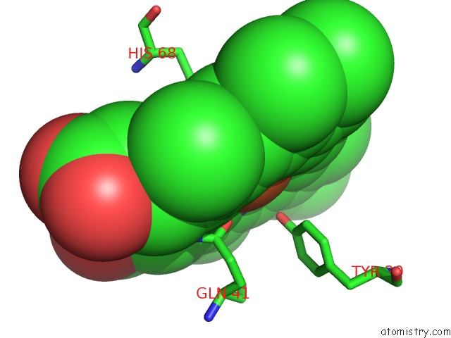
Mono view
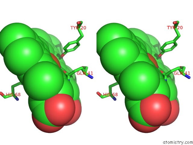
Stereo pair view
Mono view
Stereo pair view
A full contact list of Iron with other atoms in the Fe binding
site number 1 of X-Ray Crystal Structure of Truncated Hemoglobin From P.Caudatum. within 5.0Å range:
|
Reference:
A.Pesce,
M.Couture,
S.Dewilde,
M.Guertin,
K.Yamauchi,
P.Ascenzi,
L.Moens,
M.Bolognesi.
A Novel Two-Over-Two Alpha-Helical Sandwich Fold Is Characteristic of the Truncated Hemoglobin Family. Embo J. V. 19 2424 2000.
ISSN: ISSN 0261-4189
PubMed: 10835341
DOI: 10.1093/EMBOJ/19.11.2424
Page generated: Sat Aug 3 03:48:37 2024
ISSN: ISSN 0261-4189
PubMed: 10835341
DOI: 10.1093/EMBOJ/19.11.2424
Last articles
Zn in 9JYWZn in 9IR4
Zn in 9IR3
Zn in 9GMX
Zn in 9GMW
Zn in 9JEJ
Zn in 9ERF
Zn in 9ERE
Zn in 9EGV
Zn in 9EGW