Iron »
PDB 1dj7-1dry »
1do7 »
Iron in PDB 1do7: Carbonmonoxy-Myoglobin (Mutant L29W) Rebinding Structure After Photolysis at T< 180K
Protein crystallography data
The structure of Carbonmonoxy-Myoglobin (Mutant L29W) Rebinding Structure After Photolysis at T< 180K, PDB code: 1do7
was solved by
A.Ostermann,
R.Waschipky,
F.G.Parak,
G.U.Nienhaus,
with X-Ray Crystallography technique. A brief refinement statistics is given in the table below:
| Resolution Low / High (Å) | 7.00 / 1.85 |
| Space group | P 6 |
| Cell size a, b, c (Å), α, β, γ (°) | 90.430, 90.430, 45.240, 90.00, 90.00, 120.00 |
| R / Rfree (%) | 18.6 / 21.4 |
Iron Binding Sites:
The binding sites of Iron atom in the Carbonmonoxy-Myoglobin (Mutant L29W) Rebinding Structure After Photolysis at T< 180K
(pdb code 1do7). This binding sites where shown within
5.0 Angstroms radius around Iron atom.
In total only one binding site of Iron was determined in the Carbonmonoxy-Myoglobin (Mutant L29W) Rebinding Structure After Photolysis at T< 180K, PDB code: 1do7:
In total only one binding site of Iron was determined in the Carbonmonoxy-Myoglobin (Mutant L29W) Rebinding Structure After Photolysis at T< 180K, PDB code: 1do7:
Iron binding site 1 out of 1 in 1do7
Go back to
Iron binding site 1 out
of 1 in the Carbonmonoxy-Myoglobin (Mutant L29W) Rebinding Structure After Photolysis at T< 180K
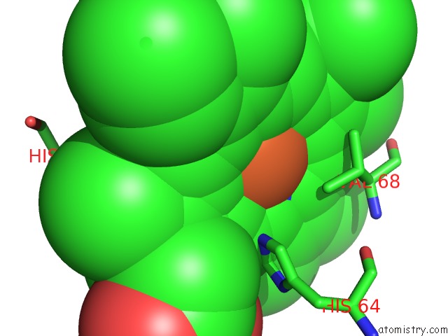
Mono view
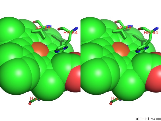
Stereo pair view
Mono view
Stereo pair view
A full contact list of Iron with other atoms in the Fe binding
site number 1 of Carbonmonoxy-Myoglobin (Mutant L29W) Rebinding Structure After Photolysis at T< 180K within 5.0Å range:
|
Reference:
A.Ostermann,
R.Waschipky,
F.G.Parak,
G.U.Nienhaus.
Ligand Binding and Conformational Motions in Myoglobin. Nature V. 404 205 2000.
ISSN: ISSN 0028-0836
PubMed: 10724176
DOI: 10.1038/35004622
Page generated: Sat Aug 3 03:53:43 2024
ISSN: ISSN 0028-0836
PubMed: 10724176
DOI: 10.1038/35004622
Last articles
Zn in 9JYWZn in 9IR4
Zn in 9IR3
Zn in 9GMX
Zn in 9GMW
Zn in 9JEJ
Zn in 9ERF
Zn in 9ERE
Zn in 9EGV
Zn in 9EGW