Iron »
PDB 1dj7-1dry »
1doi »
Iron in PDB 1doi: 2FE-2S Ferredoxin From Haloarcula Marismortui
Protein crystallography data
The structure of 2FE-2S Ferredoxin From Haloarcula Marismortui, PDB code: 1doi
was solved by
F.Frolow,
M.Harel,
J.L.Sussman,
M.Shoham,
with X-Ray Crystallography technique. A brief refinement statistics is given in the table below:
| Resolution Low / High (Å) | 30.00 / 1.90 |
| Space group | P 63 2 2 |
| Cell size a, b, c (Å), α, β, γ (°) | 59.180, 59.180, 124.230, 90.00, 90.00, 120.00 |
| R / Rfree (%) | 19.5 / n/a |
Other elements in 1doi:
The structure of 2FE-2S Ferredoxin From Haloarcula Marismortui also contains other interesting chemical elements:
| Potassium | (K) | 6 atoms |
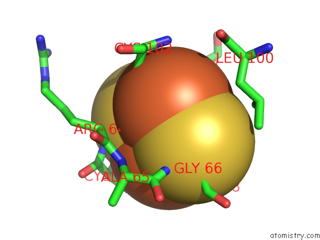
Iron Binding Sites:
The binding sites of Iron atom in the 2FE-2S Ferredoxin From Haloarcula Marismortui
(pdb code 1doi). This binding sites where shown within
5.0 Angstroms radius around Iron atom.
In total 2 binding sites of Iron where determined in the 2FE-2S Ferredoxin From Haloarcula Marismortui, PDB code: 1doi:
Jump to Iron binding site number: 1; 2;
In total 2 binding sites of Iron where determined in the 2FE-2S Ferredoxin From Haloarcula Marismortui, PDB code: 1doi:
Jump to Iron binding site number: 1; 2;
Iron binding site 1 out of 2 in 1doi
Go back to
Iron binding site 1 out
of 2 in the 2FE-2S Ferredoxin From Haloarcula Marismortui

Mono view

Stereo pair view
Mono view
Stereo pair view
A full contact list of Iron with other atoms in the Fe binding
site number 1 of 2FE-2S Ferredoxin From Haloarcula Marismortui within 5.0Å range:
|
Iron binding site 2 out of 2 in 1doi
Go back to
Iron binding site 2 out
of 2 in the 2FE-2S Ferredoxin From Haloarcula Marismortui
Mono view

Stereo pair view
Mono view
Stereo pair view
A full contact list of Iron with other atoms in the Fe binding
site number 2 of 2FE-2S Ferredoxin From Haloarcula Marismortui within 5.0Å range:
|
Reference:
F.Frolow,
M.Harel,
J.L.Sussman,
M.Mevarech,
M.Shoham.
Insights Into Protein Adaptation to A Saturated Salt Environment From the Crystal Structure of A Halophilic 2FE-2S Ferredoxin. Nat.Struct.Biol. V. 3 452 1996.
ISSN: ISSN 1072-8368
PubMed: 8612076
DOI: 10.1038/NSB0596-452
Page generated: Wed Jul 16 13:23:04 2025
ISSN: ISSN 1072-8368
PubMed: 8612076
DOI: 10.1038/NSB0596-452
Last articles
Fe in 2GP3Fe in 2GOK
Fe in 2GOJ
Fe in 2GNW
Fe in 2GNV
Fe in 2GNU
Fe in 2GMR
Fe in 2GLN
Fe in 2GL3
Fe in 2GKN