Iron »
PDB 1dj7-1dry »
1drm »
Iron in PDB 1drm: Crystal Structure of the Ligand Free Bjfixl Heme Domain
Protein crystallography data
The structure of Crystal Structure of the Ligand Free Bjfixl Heme Domain, PDB code: 1drm
was solved by
W.Gong,
B.Hao,
S.S.Mansy,
G.Gonzalez,
M.A.Gilles-Gonzalez,
M.K.Chan,
with X-Ray Crystallography technique. A brief refinement statistics is given in the table below:
| Resolution Low / High (Å) | 20.00 / 2.40 |
| Space group | H 3 2 |
| Cell size a, b, c (Å), α, β, γ (°) | 128.800, 128.800, 58.900, 90.00, 90.00, 120.00 |
| R / Rfree (%) | 19.9 / 24.8 |
Iron Binding Sites:
The binding sites of Iron atom in the Crystal Structure of the Ligand Free Bjfixl Heme Domain
(pdb code 1drm). This binding sites where shown within
5.0 Angstroms radius around Iron atom.
In total only one binding site of Iron was determined in the Crystal Structure of the Ligand Free Bjfixl Heme Domain, PDB code: 1drm:
In total only one binding site of Iron was determined in the Crystal Structure of the Ligand Free Bjfixl Heme Domain, PDB code: 1drm:
Iron binding site 1 out of 1 in 1drm
Go back to
Iron binding site 1 out
of 1 in the Crystal Structure of the Ligand Free Bjfixl Heme Domain
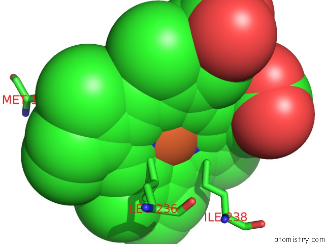
Mono view
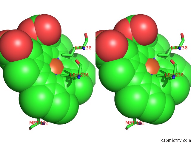
Stereo pair view
Mono view
Stereo pair view
A full contact list of Iron with other atoms in the Fe binding
site number 1 of Crystal Structure of the Ligand Free Bjfixl Heme Domain within 5.0Å range:
|
Reference:
W.Gong,
B.Hao,
S.S.Mansy,
G.Gonzalez,
M.A.Gilles-Gonzalez,
M.K.Chan.
Structure of A Biological Oxygen Sensor: A New Mechanism For Heme-Driven Signal Transduction. Proc.Natl.Acad.Sci.Usa V. 95 15177 1998.
ISSN: ISSN 0027-8424
PubMed: 9860942
DOI: 10.1073/PNAS.95.26.15177
Page generated: Sat Aug 3 03:56:14 2024
ISSN: ISSN 0027-8424
PubMed: 9860942
DOI: 10.1073/PNAS.95.26.15177
Last articles
Zn in 9JYWZn in 9IR4
Zn in 9IR3
Zn in 9GMX
Zn in 9GMW
Zn in 9JEJ
Zn in 9ERF
Zn in 9ERE
Zn in 9EGV
Zn in 9EGW