Iron in PDB 1dwl: The Ferredoxin-Cytochrome Complex Using Heteronuclear uc(Nmr) and Docking Simulation
Iron Binding Sites:
The binding sites of Iron atom in the The Ferredoxin-Cytochrome Complex Using Heteronuclear uc(Nmr) and Docking Simulation
(pdb code 1dwl). This binding sites where shown within
5.0 Angstroms radius around Iron atom.
In total 5 binding sites of Iron where determined in the
The Ferredoxin-Cytochrome Complex Using Heteronuclear uc(Nmr) and Docking Simulation, PDB code: 1dwl:
Jump to Iron binding site number:
1;
2;
3;
4;
5;
Iron binding site 1 out
of 5 in 1dwl
Go back to
Iron Binding Sites List in 1dwl
Iron binding site 1 out
of 5 in the The Ferredoxin-Cytochrome Complex Using Heteronuclear uc(Nmr) and Docking Simulation
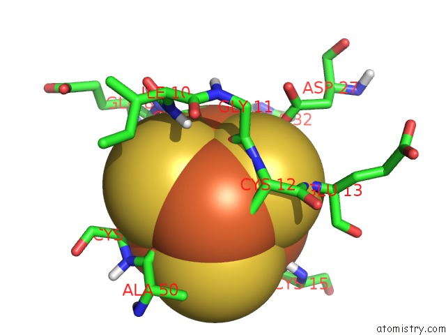
Mono view
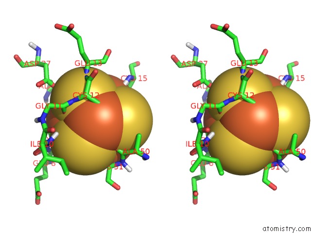
Stereo pair view
|
A full contact list of Iron with other atoms in the Fe binding
site number 1 of The Ferredoxin-Cytochrome Complex Using Heteronuclear uc(Nmr) and Docking Simulation within 5.0Å range:
probe
|
atom
|
residue
|
distance (Å)
|
B
|
Occ
|
A:Fe60
b:1.0
occ:1.00
|
FE1
|
A:SF460
|
0.0
|
1.0
|
1.0
|
S4
|
A:SF460
|
2.3
|
1.0
|
1.0
|
S3
|
A:SF460
|
2.3
|
1.0
|
1.0
|
S2
|
A:SF460
|
2.3
|
1.0
|
1.0
|
SG
|
A:CYS9
|
2.3
|
1.0
|
1.0
|
FE4
|
A:SF460
|
2.5
|
1.0
|
1.0
|
FE3
|
A:SF460
|
2.5
|
1.0
|
1.0
|
FE2
|
A:SF460
|
2.6
|
1.0
|
1.0
|
CB
|
A:CYS9
|
3.4
|
1.0
|
1.0
|
CA
|
A:CYS9
|
3.7
|
1.0
|
1.0
|
O
|
A:GLU8
|
3.8
|
1.0
|
1.0
|
S1
|
A:SF460
|
3.8
|
1.0
|
1.0
|
O
|
A:GLY11
|
3.9
|
1.0
|
1.0
|
CB
|
A:ALA32
|
4.4
|
1.0
|
1.0
|
OD1
|
A:ASP27
|
4.5
|
1.0
|
1.0
|
N
|
A:CYS9
|
4.5
|
1.0
|
1.0
|
H
|
A:ILE10
|
4.5
|
0.0
|
1.0
|
SG
|
A:CYS12
|
4.5
|
1.0
|
1.0
|
C
|
A:GLU8
|
4.6
|
1.0
|
1.0
|
H
|
A:GLU13
|
4.6
|
0.0
|
1.0
|
SG
|
A:CYS15
|
4.6
|
1.0
|
1.0
|
SG
|
A:CYS51
|
4.7
|
1.0
|
1.0
|
N
|
A:CYS12
|
4.9
|
1.0
|
1.0
|
C
|
A:CYS9
|
5.0
|
1.0
|
1.0
|
O
|
A:ALA50
|
5.0
|
1.0
|
1.0
|
|
Iron binding site 2 out
of 5 in 1dwl
Go back to
Iron Binding Sites List in 1dwl
Iron binding site 2 out
of 5 in the The Ferredoxin-Cytochrome Complex Using Heteronuclear uc(Nmr) and Docking Simulation
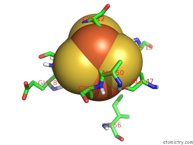
Mono view
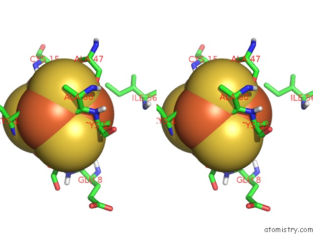
Stereo pair view
|
A full contact list of Iron with other atoms in the Fe binding
site number 2 of The Ferredoxin-Cytochrome Complex Using Heteronuclear uc(Nmr) and Docking Simulation within 5.0Å range:
probe
|
atom
|
residue
|
distance (Å)
|
B
|
Occ
|
A:Fe60
b:1.0
occ:1.00
|
FE2
|
A:SF460
|
0.0
|
1.0
|
1.0
|
S1
|
A:SF460
|
2.3
|
1.0
|
1.0
|
S3
|
A:SF460
|
2.3
|
1.0
|
1.0
|
S4
|
A:SF460
|
2.3
|
1.0
|
1.0
|
SG
|
A:CYS51
|
2.3
|
1.0
|
1.0
|
FE4
|
A:SF460
|
2.6
|
1.0
|
1.0
|
FE1
|
A:SF460
|
2.6
|
1.0
|
1.0
|
FE3
|
A:SF460
|
2.6
|
1.0
|
1.0
|
O
|
A:ALA50
|
3.4
|
1.0
|
1.0
|
CB
|
A:CYS51
|
3.6
|
1.0
|
1.0
|
CA
|
A:CYS51
|
3.7
|
1.0
|
1.0
|
O
|
A:ALA47
|
3.8
|
1.0
|
1.0
|
S2
|
A:SF460
|
3.9
|
1.0
|
1.0
|
C
|
A:ALA50
|
4.0
|
1.0
|
1.0
|
N
|
A:CYS51
|
4.0
|
1.0
|
1.0
|
CD1
|
A:ILE56
|
4.6
|
1.0
|
1.0
|
SG
|
A:CYS12
|
4.6
|
1.0
|
1.0
|
O
|
A:GLU8
|
4.7
|
1.0
|
1.0
|
SG
|
A:CYS9
|
4.7
|
1.0
|
1.0
|
SG
|
A:CYS15
|
4.7
|
1.0
|
1.0
|
H
|
A:CYS51
|
5.0
|
0.0
|
1.0
|
C
|
A:ALA47
|
5.0
|
1.0
|
1.0
|
|
Iron binding site 3 out
of 5 in 1dwl
Go back to
Iron Binding Sites List in 1dwl
Iron binding site 3 out
of 5 in the The Ferredoxin-Cytochrome Complex Using Heteronuclear uc(Nmr) and Docking Simulation
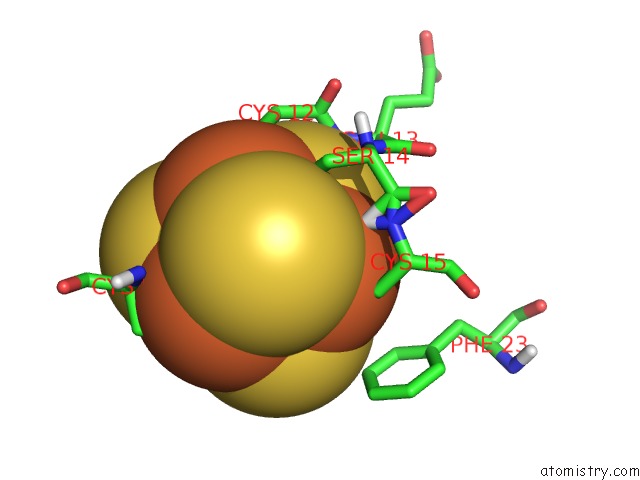
Mono view
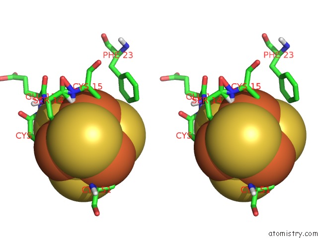
Stereo pair view
|
A full contact list of Iron with other atoms in the Fe binding
site number 3 of The Ferredoxin-Cytochrome Complex Using Heteronuclear uc(Nmr) and Docking Simulation within 5.0Å range:
probe
|
atom
|
residue
|
distance (Å)
|
B
|
Occ
|
A:Fe60
b:1.0
occ:1.00
|
FE3
|
A:SF460
|
0.0
|
1.0
|
1.0
|
S1
|
A:SF460
|
2.3
|
1.0
|
1.0
|
S4
|
A:SF460
|
2.3
|
1.0
|
1.0
|
S2
|
A:SF460
|
2.3
|
1.0
|
1.0
|
SG
|
A:CYS15
|
2.3
|
1.0
|
1.0
|
FE1
|
A:SF460
|
2.5
|
1.0
|
1.0
|
FE2
|
A:SF460
|
2.6
|
1.0
|
1.0
|
FE4
|
A:SF460
|
2.6
|
1.0
|
1.0
|
H
|
A:CYS15
|
3.5
|
0.0
|
1.0
|
CB
|
A:CYS15
|
3.6
|
1.0
|
1.0
|
OG
|
A:SER14
|
3.6
|
1.0
|
1.0
|
S3
|
A:SF460
|
3.9
|
1.0
|
1.0
|
HG
|
A:SER14
|
4.0
|
0.0
|
1.0
|
H
|
A:GLU13
|
4.0
|
0.0
|
1.0
|
N
|
A:CYS15
|
4.3
|
1.0
|
1.0
|
SG
|
A:CYS9
|
4.6
|
1.0
|
1.0
|
CA
|
A:CYS15
|
4.6
|
1.0
|
1.0
|
CB
|
A:SER14
|
4.7
|
1.0
|
1.0
|
CE2
|
A:PHE23
|
4.7
|
1.0
|
1.0
|
SG
|
A:CYS51
|
4.7
|
1.0
|
1.0
|
SG
|
A:CYS12
|
4.7
|
1.0
|
1.0
|
CB
|
A:ALA32
|
4.8
|
1.0
|
1.0
|
C
|
A:SER14
|
4.9
|
1.0
|
1.0
|
CD2
|
A:PHE23
|
4.9
|
1.0
|
1.0
|
N
|
A:SER14
|
4.9
|
1.0
|
1.0
|
|
Iron binding site 4 out
of 5 in 1dwl
Go back to
Iron Binding Sites List in 1dwl
Iron binding site 4 out
of 5 in the The Ferredoxin-Cytochrome Complex Using Heteronuclear uc(Nmr) and Docking Simulation
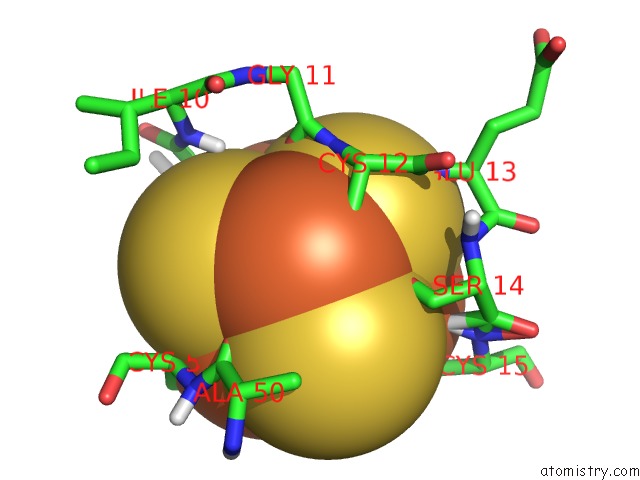
Mono view
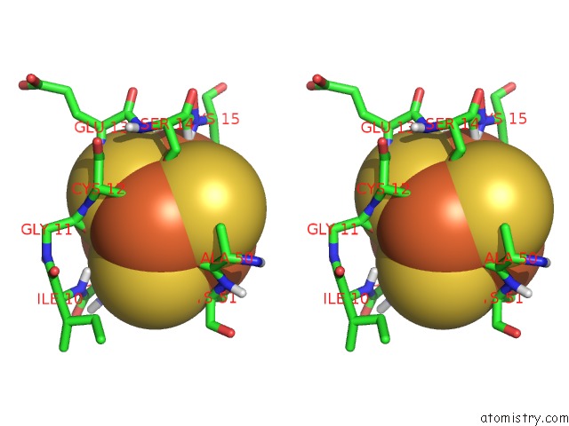
Stereo pair view
|
A full contact list of Iron with other atoms in the Fe binding
site number 4 of The Ferredoxin-Cytochrome Complex Using Heteronuclear uc(Nmr) and Docking Simulation within 5.0Å range:
probe
|
atom
|
residue
|
distance (Å)
|
B
|
Occ
|
A:Fe60
b:1.0
occ:1.00
|
FE4
|
A:SF460
|
0.0
|
1.0
|
1.0
|
SG
|
A:CYS12
|
2.3
|
1.0
|
1.0
|
S3
|
A:SF460
|
2.3
|
1.0
|
1.0
|
S1
|
A:SF460
|
2.3
|
1.0
|
1.0
|
S2
|
A:SF460
|
2.3
|
1.0
|
1.0
|
FE1
|
A:SF460
|
2.5
|
1.0
|
1.0
|
FE2
|
A:SF460
|
2.6
|
1.0
|
1.0
|
FE3
|
A:SF460
|
2.6
|
1.0
|
1.0
|
O
|
A:ALA50
|
3.1
|
1.0
|
1.0
|
OG
|
A:SER14
|
3.2
|
1.0
|
1.0
|
CB
|
A:CYS12
|
3.4
|
1.0
|
1.0
|
O
|
A:GLY11
|
3.5
|
1.0
|
1.0
|
HG
|
A:SER14
|
3.6
|
0.0
|
1.0
|
S4
|
A:SF460
|
3.9
|
1.0
|
1.0
|
N
|
A:CYS12
|
3.9
|
1.0
|
1.0
|
H
|
A:GLU13
|
3.9
|
0.0
|
1.0
|
CA
|
A:CYS12
|
4.2
|
1.0
|
1.0
|
C
|
A:ALA50
|
4.2
|
1.0
|
1.0
|
C
|
A:GLY11
|
4.4
|
1.0
|
1.0
|
H
|
A:ILE10
|
4.5
|
0.0
|
1.0
|
CB
|
A:SER14
|
4.5
|
1.0
|
1.0
|
SG
|
A:CYS9
|
4.6
|
1.0
|
1.0
|
SG
|
A:CYS51
|
4.7
|
1.0
|
1.0
|
N
|
A:GLU13
|
4.7
|
1.0
|
1.0
|
H
|
A:CYS15
|
4.7
|
0.0
|
1.0
|
SG
|
A:CYS15
|
4.7
|
1.0
|
1.0
|
CB
|
A:ALA50
|
4.7
|
1.0
|
1.0
|
C
|
A:CYS12
|
4.8
|
1.0
|
1.0
|
CA
|
A:CYS51
|
4.8
|
1.0
|
1.0
|
N
|
A:CYS51
|
4.9
|
1.0
|
1.0
|
CB
|
A:CYS9
|
4.9
|
1.0
|
1.0
|
CA
|
A:CYS9
|
4.9
|
1.0
|
1.0
|
|
Iron binding site 5 out
of 5 in 1dwl
Go back to
Iron Binding Sites List in 1dwl
Iron binding site 5 out
of 5 in the The Ferredoxin-Cytochrome Complex Using Heteronuclear uc(Nmr) and Docking Simulation
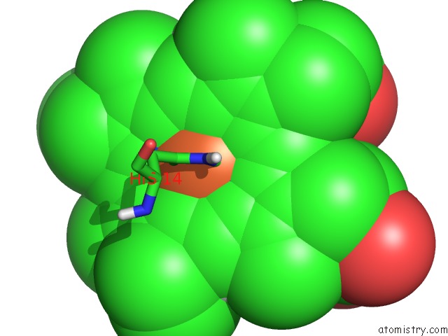
Mono view
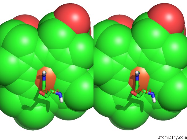
Stereo pair view
|
A full contact list of Iron with other atoms in the Fe binding
site number 5 of The Ferredoxin-Cytochrome Complex Using Heteronuclear uc(Nmr) and Docking Simulation within 5.0Å range:
probe
|
atom
|
residue
|
distance (Å)
|
B
|
Occ
|
B:Fe80
b:1.0
occ:1.00
|
FE
|
B:HEC80
|
0.0
|
1.0
|
1.0
|
NA
|
B:HEC80
|
2.0
|
1.0
|
1.0
|
NC
|
B:HEC80
|
2.0
|
1.0
|
1.0
|
ND
|
B:HEC80
|
2.0
|
1.0
|
1.0
|
NB
|
B:HEC80
|
2.0
|
1.0
|
1.0
|
NE2
|
B:HIS14
|
2.2
|
1.0
|
1.0
|
SD
|
B:MET57
|
2.3
|
1.0
|
1.0
|
C1C
|
B:HEC80
|
3.0
|
1.0
|
1.0
|
C4A
|
B:HEC80
|
3.0
|
1.0
|
1.0
|
C1D
|
B:HEC80
|
3.0
|
1.0
|
1.0
|
C4C
|
B:HEC80
|
3.0
|
1.0
|
1.0
|
C1B
|
B:HEC80
|
3.0
|
1.0
|
1.0
|
C1A
|
B:HEC80
|
3.0
|
1.0
|
1.0
|
C4D
|
B:HEC80
|
3.0
|
1.0
|
1.0
|
C4B
|
B:HEC80
|
3.0
|
1.0
|
1.0
|
CE1
|
B:HIS14
|
3.2
|
1.0
|
1.0
|
CD2
|
B:HIS14
|
3.2
|
1.0
|
1.0
|
CHC
|
B:HEC80
|
3.4
|
1.0
|
1.0
|
CHB
|
B:HEC80
|
3.4
|
1.0
|
1.0
|
CHD
|
B:HEC80
|
3.4
|
1.0
|
1.0
|
CHA
|
B:HEC80
|
3.4
|
1.0
|
1.0
|
CE
|
B:MET57
|
3.6
|
1.0
|
1.0
|
CG
|
B:MET57
|
3.6
|
1.0
|
1.0
|
C2C
|
B:HEC80
|
4.2
|
1.0
|
1.0
|
C3A
|
B:HEC80
|
4.2
|
1.0
|
1.0
|
C2B
|
B:HEC80
|
4.2
|
1.0
|
1.0
|
C2D
|
B:HEC80
|
4.2
|
1.0
|
1.0
|
C2A
|
B:HEC80
|
4.2
|
1.0
|
1.0
|
C3D
|
B:HEC80
|
4.2
|
1.0
|
1.0
|
C3C
|
B:HEC80
|
4.3
|
1.0
|
1.0
|
C3B
|
B:HEC80
|
4.3
|
1.0
|
1.0
|
ND1
|
B:HIS14
|
4.3
|
1.0
|
1.0
|
CG
|
B:HIS14
|
4.3
|
1.0
|
1.0
|
HHC
|
B:HEC80
|
4.4
|
0.0
|
1.0
|
HHD
|
B:HEC80
|
4.5
|
0.0
|
1.0
|
HHB
|
B:HEC80
|
4.5
|
0.0
|
1.0
|
HHA
|
B:HEC80
|
4.5
|
0.0
|
1.0
|
CB
|
B:MET57
|
5.0
|
1.0
|
1.0
|
|
Reference:
X.Morelli,
A.Dolla,
M.Czjzek,
P.N.Palma,
F.Blasco,
L.Krippahl,
J.J.G.Moura,
F.Guerlesquin.
Heteronuclear uc(Nmr) and Soft Docking: An Experimental Approach For A Structural Model of the Cytochrome C553-Ferredoxin Complex Biochemistry V. 39 2530 2000.
ISSN: ISSN 0006-2960
PubMed: 10704202
DOI: 10.1021/BI992306S
Page generated: Sat Aug 3 04:02:15 2024
|