Iron »
PDB 1dxt-1ea1 »
1dyt »
Iron in PDB 1dyt: X-Ray Crystal Structure of Ecp (Rnase 3) at 1.75 A
Protein crystallography data
The structure of X-Ray Crystal Structure of Ecp (Rnase 3) at 1.75 A, PDB code: 1dyt
was solved by
G.Mallorqui-Fernandez,
J.Pous,
R.Peracaula,
T.Maeda,
H.Tada,
H.Yamada,
M.Seno,
R.De Llorens,
F.X.Gomis-Rueth,
M.Coll,
with X-Ray Crystallography technique. A brief refinement statistics is given in the table below:
| Resolution Low / High (Å) | 20 / 1.75 |
| Space group | P 43 2 2 |
| Cell size a, b, c (Å), α, β, γ (°) | 62.163, 62.163, 174.590, 90.00, 90.00, 90.00 |
| R / Rfree (%) | 22.4 / 27.1 |
Iron Binding Sites:
The binding sites of Iron atom in the X-Ray Crystal Structure of Ecp (Rnase 3) at 1.75 A
(pdb code 1dyt). This binding sites where shown within
5.0 Angstroms radius around Iron atom.
In total 2 binding sites of Iron where determined in the X-Ray Crystal Structure of Ecp (Rnase 3) at 1.75 A, PDB code: 1dyt:
Jump to Iron binding site number: 1; 2;
In total 2 binding sites of Iron where determined in the X-Ray Crystal Structure of Ecp (Rnase 3) at 1.75 A, PDB code: 1dyt:
Jump to Iron binding site number: 1; 2;
Iron binding site 1 out of 2 in 1dyt
Go back to
Iron binding site 1 out
of 2 in the X-Ray Crystal Structure of Ecp (Rnase 3) at 1.75 A
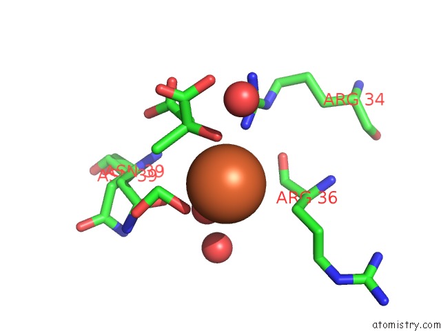
Mono view
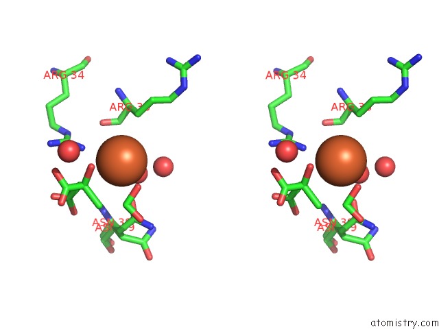
Stereo pair view
Mono view
Stereo pair view
A full contact list of Iron with other atoms in the Fe binding
site number 1 of X-Ray Crystal Structure of Ecp (Rnase 3) at 1.75 A within 5.0Å range:
|
Iron binding site 2 out of 2 in 1dyt
Go back to
Iron binding site 2 out
of 2 in the X-Ray Crystal Structure of Ecp (Rnase 3) at 1.75 A
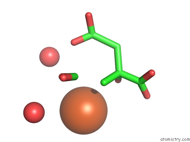
Mono view
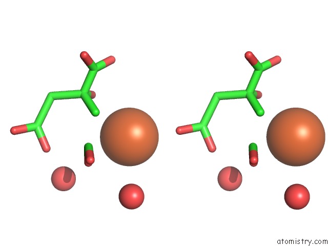
Stereo pair view
Mono view
Stereo pair view
A full contact list of Iron with other atoms in the Fe binding
site number 2 of X-Ray Crystal Structure of Ecp (Rnase 3) at 1.75 A within 5.0Å range:
|
Reference:
G.Mallorqui-Fernandez,
J.Pous,
R.Peracaula,
T.Maeda,
H.Tada,
H.Yamada,
M.Seno,
R.De Llorens,
F.X.Gomis-Rueth,
M.Coll.
Three-Dimensional Crystal Structure of Human Eosinophil Cationic Protein (Rnase 3) at 1.75 A Resolution. J.Mol.Biol. V. 300 1297 2000.
ISSN: ISSN 0022-2836
PubMed: 10903870
DOI: 10.1006/JMBI.2000.3939
Page generated: Wed Jul 16 13:32:23 2025
ISSN: ISSN 0022-2836
PubMed: 10903870
DOI: 10.1006/JMBI.2000.3939
Last articles
Fe in 1R1YFe in 1R1X
Fe in 1R0Q
Fe in 1QYZ
Fe in 1QXE
Fe in 1QYB
Fe in 1QWS
Fe in 1QXD
Fe in 1QWM
Fe in 1QWL