Iron »
PDB 1dxt-1ea1 »
1e5s »
Iron in PDB 1e5s: Proline 3-Hydroxylase (Type II) - Iron Form
Protein crystallography data
The structure of Proline 3-Hydroxylase (Type II) - Iron Form, PDB code: 1e5s
was solved by
I.J.Clifton,
L.C.Hsueh,
J.E.Baldwin,
C.J.Schofield,
K.Harlos,
with X-Ray Crystallography technique. A brief refinement statistics is given in the table below:
| Resolution Low / High (Å) | 23.82 / 2.40 |
| Space group | P 31 2 1 |
| Cell size a, b, c (Å), α, β, γ (°) | 72.780, 72.780, 224.900, 90.00, 90.00, 120.00 |
| R / Rfree (%) | 21.6 / 27.1 |
Iron Binding Sites:
The binding sites of Iron atom in the Proline 3-Hydroxylase (Type II) - Iron Form
(pdb code 1e5s). This binding sites where shown within
5.0 Angstroms radius around Iron atom.
In total 2 binding sites of Iron where determined in the Proline 3-Hydroxylase (Type II) - Iron Form, PDB code: 1e5s:
Jump to Iron binding site number: 1; 2;
In total 2 binding sites of Iron where determined in the Proline 3-Hydroxylase (Type II) - Iron Form, PDB code: 1e5s:
Jump to Iron binding site number: 1; 2;
Iron binding site 1 out of 2 in 1e5s
Go back to
Iron binding site 1 out
of 2 in the Proline 3-Hydroxylase (Type II) - Iron Form
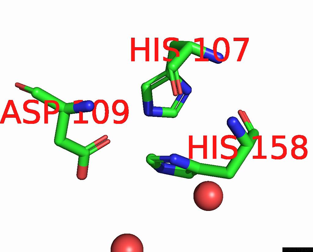
Mono view
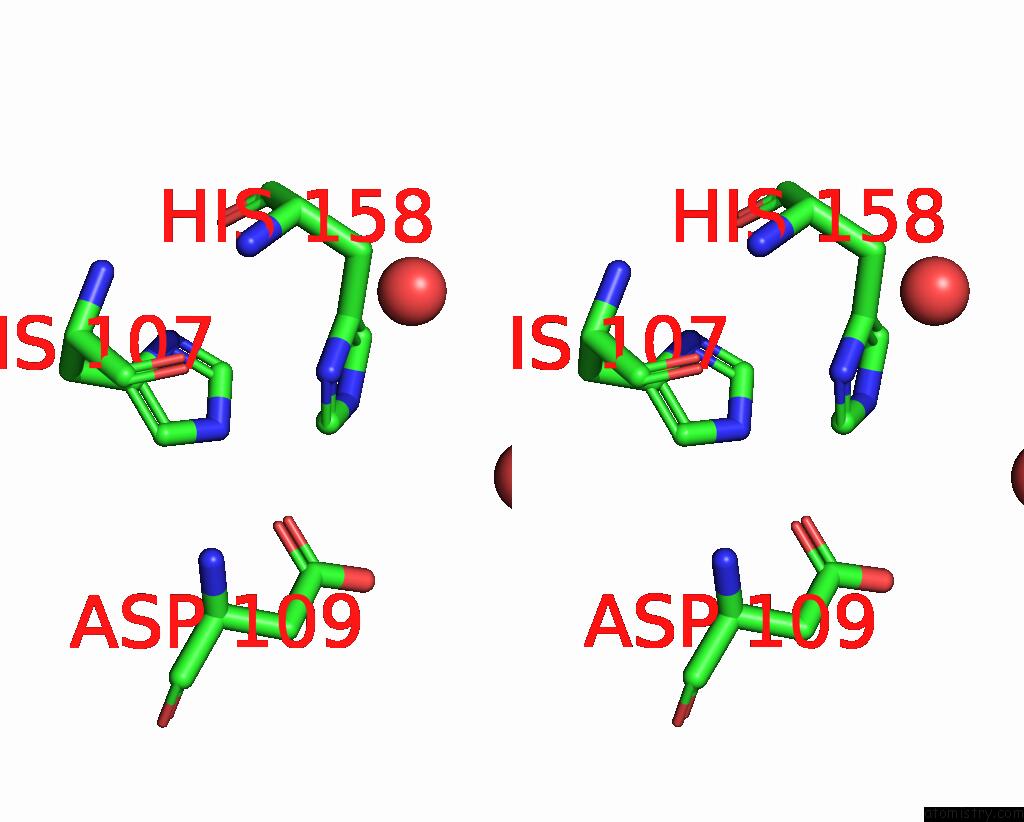
Stereo pair view
Mono view
Stereo pair view
A full contact list of Iron with other atoms in the Fe binding
site number 1 of Proline 3-Hydroxylase (Type II) - Iron Form within 5.0Å range:
|
Iron binding site 2 out of 2 in 1e5s
Go back to
Iron binding site 2 out
of 2 in the Proline 3-Hydroxylase (Type II) - Iron Form
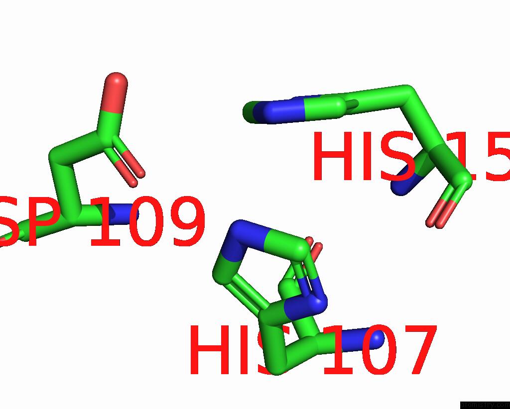
Mono view
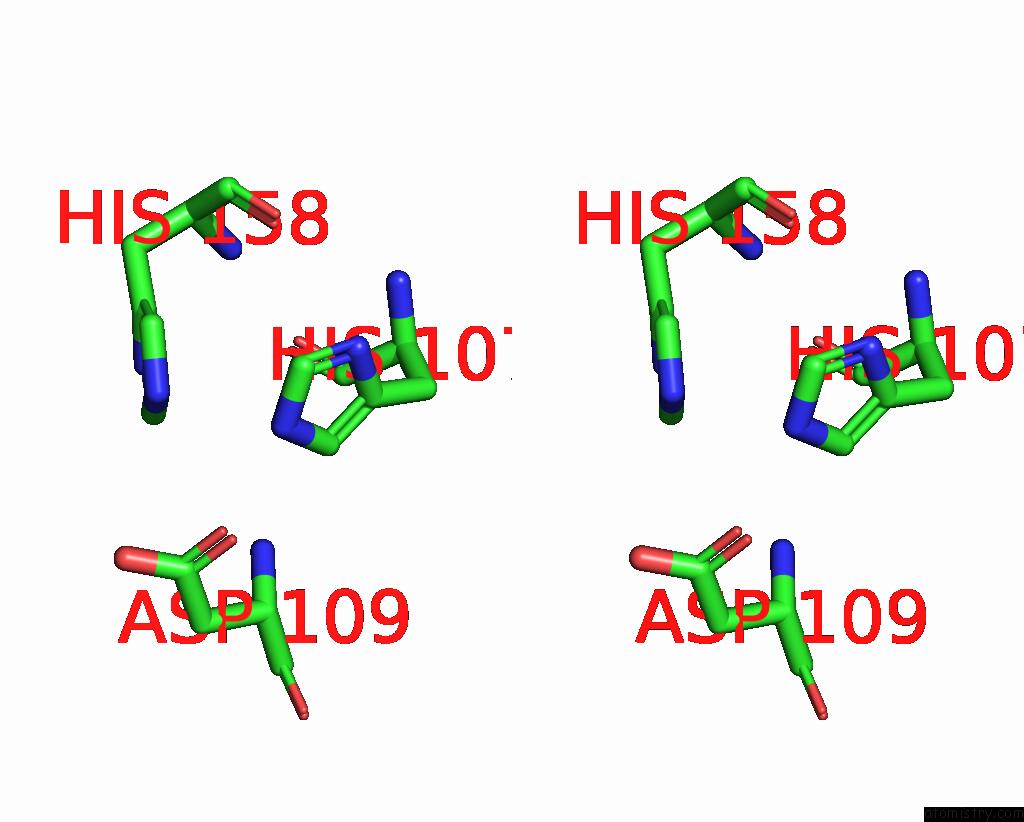
Stereo pair view
Mono view
Stereo pair view
A full contact list of Iron with other atoms in the Fe binding
site number 2 of Proline 3-Hydroxylase (Type II) - Iron Form within 5.0Å range:
|
Reference:
I.J.Clifton,
L.C.Hsueh,
J.E.Baldwin,
K.Harlos,
C.J.Schofield.
Structure of Proline 3-Hydroxylase. Evolution of the Family of 2-Oxoglutarate Dependent Oxygenases. Eur.J.Biochem. V. 268 6625 2001.
ISSN: ISSN 0014-2956
PubMed: 11737217
DOI: 10.1046/J.0014-2956.2001.02617.X
Page generated: Sat Aug 3 04:15:07 2024
ISSN: ISSN 0014-2956
PubMed: 11737217
DOI: 10.1046/J.0014-2956.2001.02617.X
Last articles
F in 4NG5F in 4NH9
F in 4NH7
F in 4NCJ
F in 4NH8
F in 4NFH
F in 4NFS
F in 4NCR
F in 4NCM
F in 4NCS