Iron »
PDB 1dxt-1ea1 »
1e9m »
Iron in PDB 1e9m: Ferredoxin VI From Rhodobacter Capsulatus
Protein crystallography data
The structure of Ferredoxin VI From Rhodobacter Capsulatus, PDB code: 1e9m
was solved by
G.Sainz,
J.Armengaud,
V.Stojanoff,
N.Sanishvili,
Y.Jouanneau,
S.Larry,
with X-Ray Crystallography technique. A brief refinement statistics is given in the table below:
| Resolution Low / High (Å) | 19.27 / 2.07 |
| Space group | P 21 21 21 |
| Cell size a, b, c (Å), α, β, γ (°) | 45.340, 49.030, 54.910, 90.00, 90.00, 90.00 |
| R / Rfree (%) | 19.6 / 21.4 |
Iron Binding Sites:
The binding sites of Iron atom in the Ferredoxin VI From Rhodobacter Capsulatus
(pdb code 1e9m). This binding sites where shown within
5.0 Angstroms radius around Iron atom.
In total 2 binding sites of Iron where determined in the Ferredoxin VI From Rhodobacter Capsulatus, PDB code: 1e9m:
Jump to Iron binding site number: 1; 2;
In total 2 binding sites of Iron where determined in the Ferredoxin VI From Rhodobacter Capsulatus, PDB code: 1e9m:
Jump to Iron binding site number: 1; 2;
Iron binding site 1 out of 2 in 1e9m
Go back to
Iron binding site 1 out
of 2 in the Ferredoxin VI From Rhodobacter Capsulatus
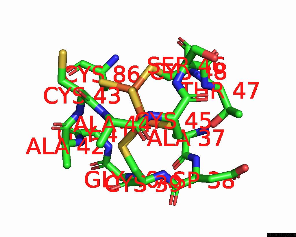
Mono view
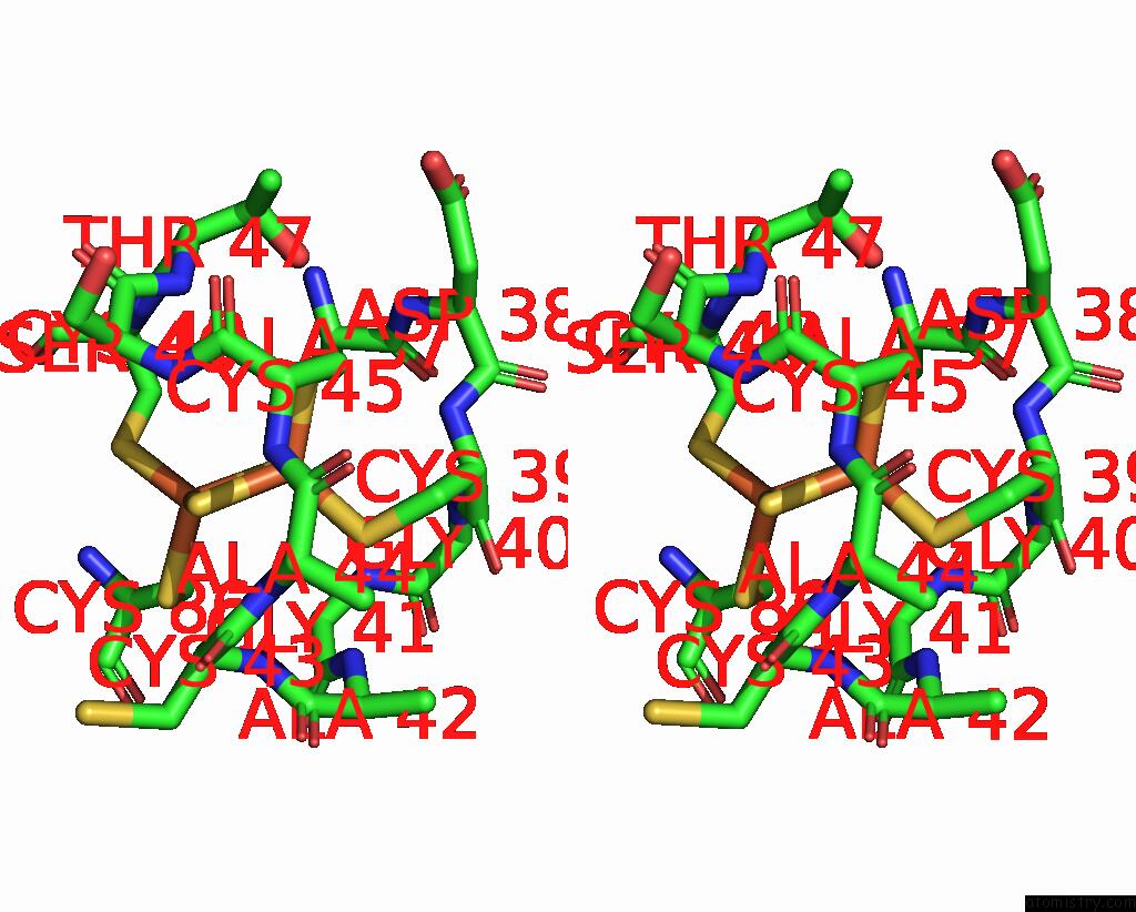
Stereo pair view
Mono view
Stereo pair view
A full contact list of Iron with other atoms in the Fe binding
site number 1 of Ferredoxin VI From Rhodobacter Capsulatus within 5.0Å range:
|
Iron binding site 2 out of 2 in 1e9m
Go back to
Iron binding site 2 out
of 2 in the Ferredoxin VI From Rhodobacter Capsulatus
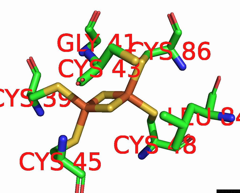
Mono view
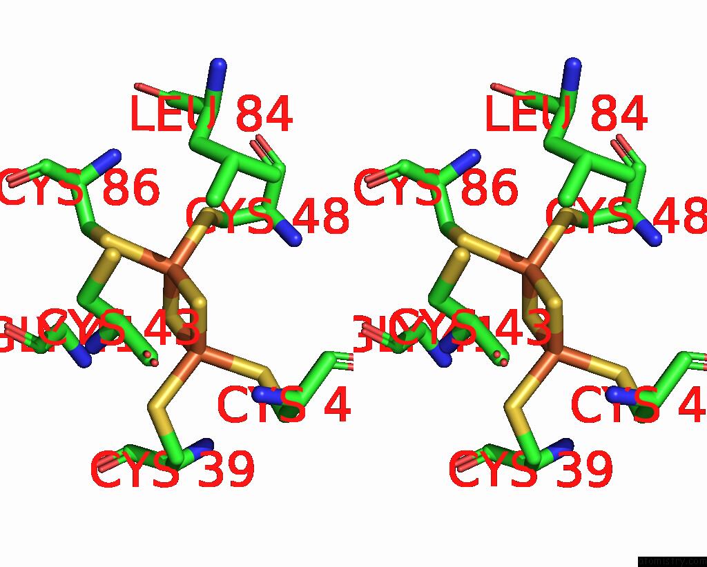
Stereo pair view
Mono view
Stereo pair view
A full contact list of Iron with other atoms in the Fe binding
site number 2 of Ferredoxin VI From Rhodobacter Capsulatus within 5.0Å range:
|
Reference:
J.Armengaud,
G.Sainz,
Y.Jouanneau,
L.C.Sieker.
Crystallization and Preliminary X-Ray Diffraction Analysis of A [2FE-2S] Ferredoxin (Fdvi) From Rhodobacter Capsulatus Acta Crystallogr.,Sect.D V. 57 301 2001.
ISSN: ISSN 0907-4449
PubMed: 11173487
DOI: 10.1107/S0907444900017832
Page generated: Sat Aug 3 04:17:22 2024
ISSN: ISSN 0907-4449
PubMed: 11173487
DOI: 10.1107/S0907444900017832
Last articles
F in 7NBLF in 7N93
F in 7NBK
F in 7NAJ
F in 7N91
F in 7N8R
F in 7N77
F in 7N7W
F in 7N73
F in 7N7L