Iron »
PDB 1dxt-1ea1 »
1e9x »
Iron in PDB 1e9x: Cytochrome P450 14 Alpha-Sterol Demethylase (CYP51) From Mycobacterium Tuberculosis in Complex with 4-Phenylimidazole
Protein crystallography data
The structure of Cytochrome P450 14 Alpha-Sterol Demethylase (CYP51) From Mycobacterium Tuberculosis in Complex with 4-Phenylimidazole, PDB code: 1e9x
was solved by
L.M.Podust,
T.L.Poulos,
M.R.Waterman,
with X-Ray Crystallography technique. A brief refinement statistics is given in the table below:
| Resolution Low / High (Å) | 39.16 / 2.10 |
| Space group | P 21 21 21 |
| Cell size a, b, c (Å), α, β, γ (°) | 46.136, 83.863, 109.557, 90.00, 90.00, 90.00 |
| R / Rfree (%) | 18.5 / 23 |
Iron Binding Sites:
The binding sites of Iron atom in the Cytochrome P450 14 Alpha-Sterol Demethylase (CYP51) From Mycobacterium Tuberculosis in Complex with 4-Phenylimidazole
(pdb code 1e9x). This binding sites where shown within
5.0 Angstroms radius around Iron atom.
In total only one binding site of Iron was determined in the Cytochrome P450 14 Alpha-Sterol Demethylase (CYP51) From Mycobacterium Tuberculosis in Complex with 4-Phenylimidazole, PDB code: 1e9x:
In total only one binding site of Iron was determined in the Cytochrome P450 14 Alpha-Sterol Demethylase (CYP51) From Mycobacterium Tuberculosis in Complex with 4-Phenylimidazole, PDB code: 1e9x:
Iron binding site 1 out of 1 in 1e9x
Go back to
Iron binding site 1 out
of 1 in the Cytochrome P450 14 Alpha-Sterol Demethylase (CYP51) From Mycobacterium Tuberculosis in Complex with 4-Phenylimidazole
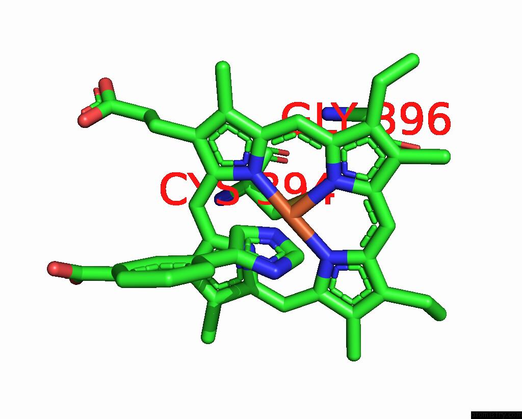
Mono view
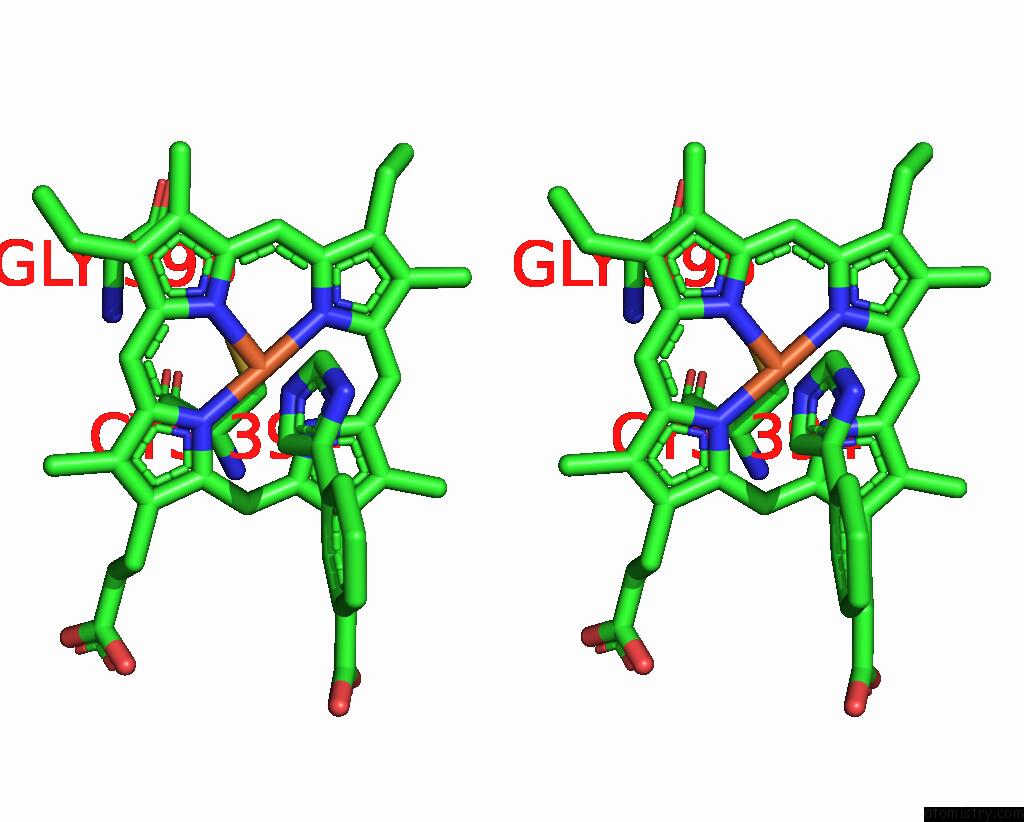
Stereo pair view
Mono view
Stereo pair view
A full contact list of Iron with other atoms in the Fe binding
site number 1 of Cytochrome P450 14 Alpha-Sterol Demethylase (CYP51) From Mycobacterium Tuberculosis in Complex with 4-Phenylimidazole within 5.0Å range:
|
Reference:
L.M.Podust,
T.L.Poulos,
M.R.Waterman.
Crystal Structure of Cytochrome P450 14ALPHA -Sterol Demethylase (CYP51) From Mycobacterium Tuberculosis in Complex with Azole Inhibitors Proc.Natl.Acad.Sci.Usa V. 98 3068 2001.
ISSN: ISSN 0027-8424
PubMed: 11248033
DOI: 10.1073/PNAS.061562898
Page generated: Wed Jul 16 13:41:20 2025
ISSN: ISSN 0027-8424
PubMed: 11248033
DOI: 10.1073/PNAS.061562898
Last articles
Fe in 2YXOFe in 2YRS
Fe in 2YXC
Fe in 2YNM
Fe in 2YVJ
Fe in 2YP1
Fe in 2YU2
Fe in 2YU1
Fe in 2YQB
Fe in 2YOO