Iron »
PDB 1dxt-1ea1 »
1ea1 »
Iron in PDB 1ea1: Cytochrome P450 14 Alpha-Sterol Demethylase (CYP51) From Mycobacterium Tuberculosis in Complex with Fluconazole
Protein crystallography data
The structure of Cytochrome P450 14 Alpha-Sterol Demethylase (CYP51) From Mycobacterium Tuberculosis in Complex with Fluconazole, PDB code: 1ea1
was solved by
L.M.Podust,
T.L.Poulos,
M.R.Waterman,
with X-Ray Crystallography technique. A brief refinement statistics is given in the table below:
| Resolution Low / High (Å) | 38.00 / 2.21 |
| Space group | P 21 21 21 |
| Cell size a, b, c (Å), α, β, γ (°) | 46.186, 84.258, 109.754, 90.00, 90.00, 90.00 |
| R / Rfree (%) | 20.4 / 24.9 |
Other elements in 1ea1:
The structure of Cytochrome P450 14 Alpha-Sterol Demethylase (CYP51) From Mycobacterium Tuberculosis in Complex with Fluconazole also contains other interesting chemical elements:
| Fluorine | (F) | 2 atoms |
Iron Binding Sites:
The binding sites of Iron atom in the Cytochrome P450 14 Alpha-Sterol Demethylase (CYP51) From Mycobacterium Tuberculosis in Complex with Fluconazole
(pdb code 1ea1). This binding sites where shown within
5.0 Angstroms radius around Iron atom.
In total only one binding site of Iron was determined in the Cytochrome P450 14 Alpha-Sterol Demethylase (CYP51) From Mycobacterium Tuberculosis in Complex with Fluconazole, PDB code: 1ea1:
In total only one binding site of Iron was determined in the Cytochrome P450 14 Alpha-Sterol Demethylase (CYP51) From Mycobacterium Tuberculosis in Complex with Fluconazole, PDB code: 1ea1:
Iron binding site 1 out of 1 in 1ea1
Go back to
Iron binding site 1 out
of 1 in the Cytochrome P450 14 Alpha-Sterol Demethylase (CYP51) From Mycobacterium Tuberculosis in Complex with Fluconazole
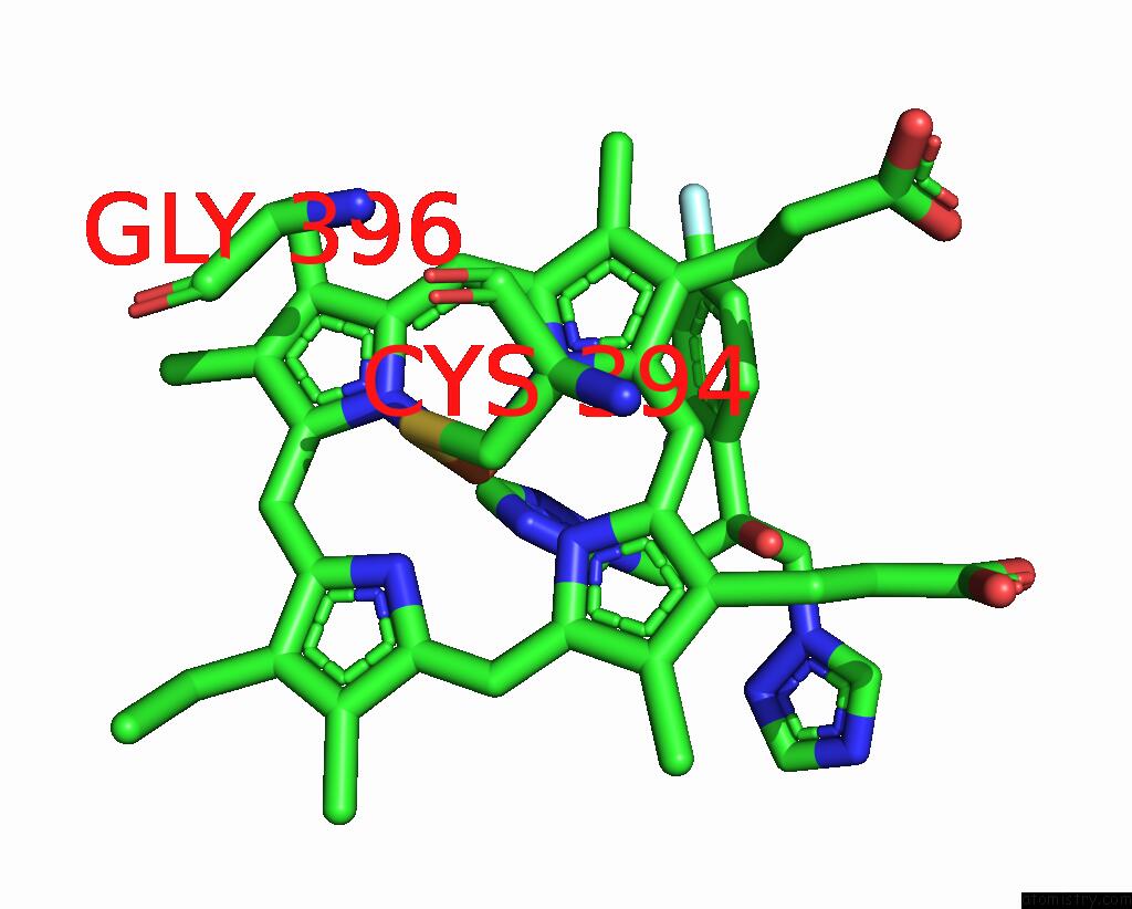
Mono view
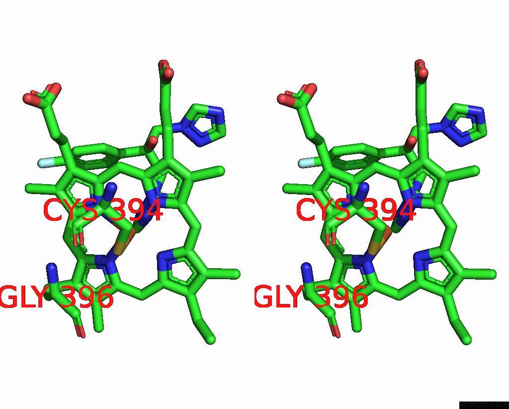
Stereo pair view
Mono view
Stereo pair view
A full contact list of Iron with other atoms in the Fe binding
site number 1 of Cytochrome P450 14 Alpha-Sterol Demethylase (CYP51) From Mycobacterium Tuberculosis in Complex with Fluconazole within 5.0Å range:
|
Reference:
L.M.Podust,
T.L.Poulos,
M.R.Waterman.
Crystal Structure of Cytochrome P450 14ALPHA -Sterol Demethylase (CYP51) From Mycobacterium Tuberculosis in Complex with Azole Inhibitors Proc.Natl.Acad.Sci.Usa V. 98 3068 2001.
ISSN: ISSN 0027-8424
PubMed: 11248033
DOI: 10.1073/PNAS.061562898
Page generated: Sat Aug 3 04:18:56 2024
ISSN: ISSN 0027-8424
PubMed: 11248033
DOI: 10.1073/PNAS.061562898
Last articles
Zn in 9J0NZn in 9J0O
Zn in 9J0P
Zn in 9FJX
Zn in 9EKB
Zn in 9C0F
Zn in 9CAH
Zn in 9CH0
Zn in 9CH3
Zn in 9CH1