Iron »
PDB 1etp-1faw »
1ew0 »
Iron in PDB 1ew0: Crystal Structure Analysis of the Sensor Domain of Rmfixl(Ferrous Form)
Protein crystallography data
The structure of Crystal Structure Analysis of the Sensor Domain of Rmfixl(Ferrous Form), PDB code: 1ew0
was solved by
H.Miyatake,
M.Mukai,
S.-Y.Park,
S.Adachi,
K.Tamura,
H.Nakamura,
K.Nakamura,
T.Tsuchiya,
T.Iizuka,
Y.Shiro,
with X-Ray Crystallography technique. A brief refinement statistics is given in the table below:
| Resolution Low / High (Å) | 20.00 / 1.40 |
| Space group | C 1 2 1 |
| Cell size a, b, c (Å), α, β, γ (°) | 60.300, 37.040, 53.450, 90.00, 115.44, 90.00 |
| R / Rfree (%) | 20.5 / 26.5 |
Iron Binding Sites:
The binding sites of Iron atom in the Crystal Structure Analysis of the Sensor Domain of Rmfixl(Ferrous Form)
(pdb code 1ew0). This binding sites where shown within
5.0 Angstroms radius around Iron atom.
In total only one binding site of Iron was determined in the Crystal Structure Analysis of the Sensor Domain of Rmfixl(Ferrous Form), PDB code: 1ew0:
In total only one binding site of Iron was determined in the Crystal Structure Analysis of the Sensor Domain of Rmfixl(Ferrous Form), PDB code: 1ew0:
Iron binding site 1 out of 1 in 1ew0
Go back to
Iron binding site 1 out
of 1 in the Crystal Structure Analysis of the Sensor Domain of Rmfixl(Ferrous Form)
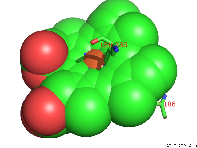
Mono view
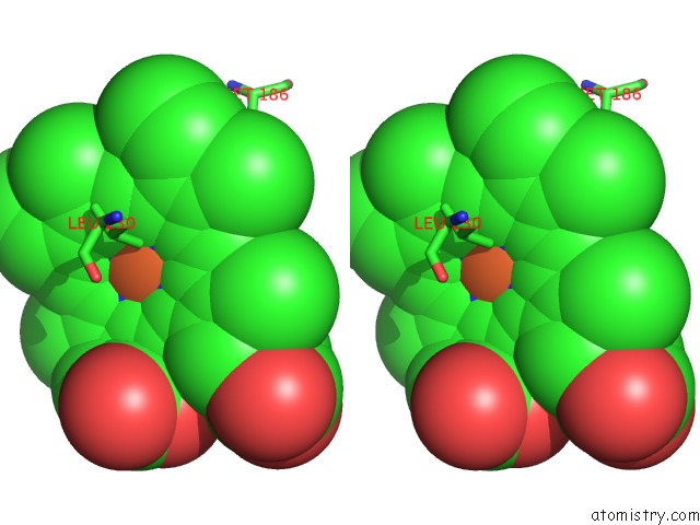
Stereo pair view
Mono view
Stereo pair view
A full contact list of Iron with other atoms in the Fe binding
site number 1 of Crystal Structure Analysis of the Sensor Domain of Rmfixl(Ferrous Form) within 5.0Å range:
|
Reference:
H.Miyatake,
M.Mukai,
S.-Y.Park,
S.Adachi,
K.Tamura,
H.Nakamura,
K.Nakamura,
T.Tsuchiya,
T.Iizuka,
Y.Shiro.
Sensory Mechanism of Oxygen Sensor Fixl From Rhizobium Meliloti: Crystallographic, Mutagenesis and Resonance Raman Spectroscopic Studies J.Mol.Biol. V. 301 415 2000.
ISSN: ISSN 0022-2836
PubMed: 10926518
DOI: 10.1006/JMBI.2000.3954
Page generated: Sat Aug 3 04:37:53 2024
ISSN: ISSN 0022-2836
PubMed: 10926518
DOI: 10.1006/JMBI.2000.3954
Last articles
Zn in 9MJ5Zn in 9HNW
Zn in 9G0L
Zn in 9FNE
Zn in 9DZN
Zn in 9E0I
Zn in 9D32
Zn in 9DAK
Zn in 8ZXC
Zn in 8ZUF