Iron »
PDB 1etp-1faw »
1f1f »
Iron in PDB 1f1f: Crystal Structure of Cytochrome C6 From Arthrospira Maxima
Protein crystallography data
The structure of Crystal Structure of Cytochrome C6 From Arthrospira Maxima, PDB code: 1f1f
was solved by
C.A.Kerfeld,
A.A.Serag,
M.R.Sawaya,
D.W.Krogmann,
T.O.Yeates,
with X-Ray Crystallography technique. A brief refinement statistics is given in the table below:
| Resolution Low / High (Å) | 50.00 / 2.70 |
| Space group | P 21 21 21 |
| Cell size a, b, c (Å), α, β, γ (°) | 79.400, 67.800, 49.700, 90.00, 90.00, 90.00 |
| R / Rfree (%) | 23.2 / 25.5 |
Iron Binding Sites:
The binding sites of Iron atom in the Crystal Structure of Cytochrome C6 From Arthrospira Maxima
(pdb code 1f1f). This binding sites where shown within
5.0 Angstroms radius around Iron atom.
In total only one binding site of Iron was determined in the Crystal Structure of Cytochrome C6 From Arthrospira Maxima, PDB code: 1f1f:
In total only one binding site of Iron was determined in the Crystal Structure of Cytochrome C6 From Arthrospira Maxima, PDB code: 1f1f:
Iron binding site 1 out of 1 in 1f1f
Go back to
Iron binding site 1 out
of 1 in the Crystal Structure of Cytochrome C6 From Arthrospira Maxima
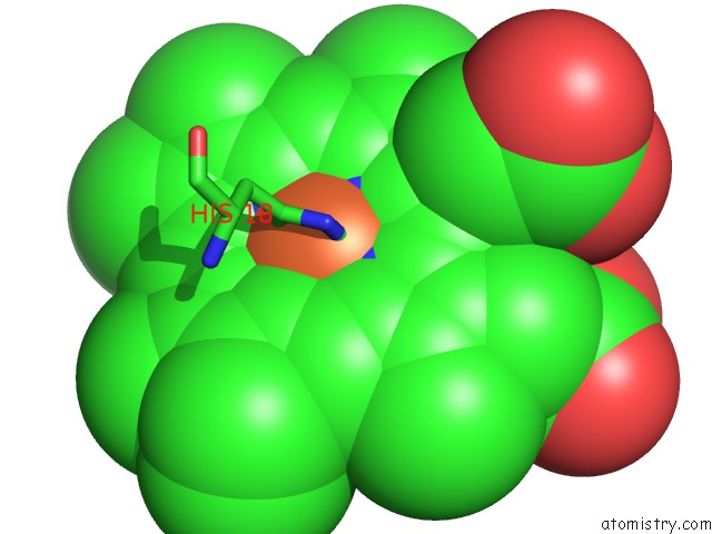
Mono view
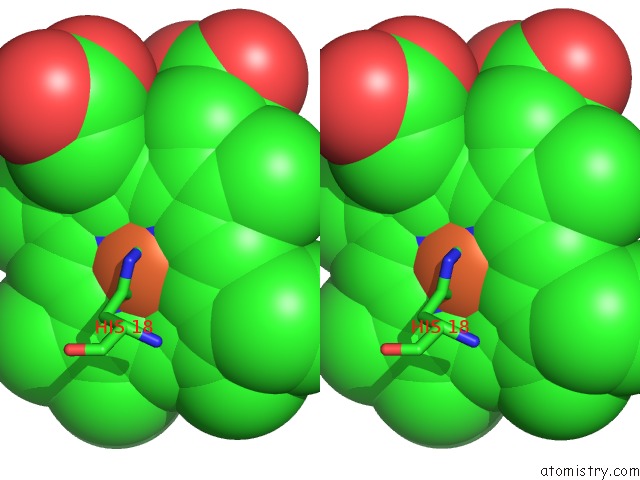
Stereo pair view
Mono view
Stereo pair view
A full contact list of Iron with other atoms in the Fe binding
site number 1 of Crystal Structure of Cytochrome C6 From Arthrospira Maxima within 5.0Å range:
|
Reference:
M.R.Sawaya,
D.W.Krogmann,
A.Serag,
K.K.Ho,
T.O.Yeates,
C.A.Kerfeld.
Structures of Cytochrome C-549 and Cytochrome C6 From the Cyanobacterium Arthrospira Maxima. Biochemistry V. 40 9215 2001.
ISSN: ISSN 0006-2960
PubMed: 11478889
DOI: 10.1021/BI002679P
Page generated: Sat Aug 3 04:40:26 2024
ISSN: ISSN 0006-2960
PubMed: 11478889
DOI: 10.1021/BI002679P
Last articles
Zn in 9J0NZn in 9J0O
Zn in 9J0P
Zn in 9FJX
Zn in 9EKB
Zn in 9C0F
Zn in 9CAH
Zn in 9CH0
Zn in 9CH3
Zn in 9CH1