Iron »
PDB 1etp-1faw »
1f5c »
Iron in PDB 1f5c: Crystal Structure of F25H Ferredoxin 1 Mutant From Azotobacter Vinelandii at 1.75 Angstrom Resolution
Protein crystallography data
The structure of Crystal Structure of F25H Ferredoxin 1 Mutant From Azotobacter Vinelandii at 1.75 Angstrom Resolution, PDB code: 1f5c
was solved by
K.Chen,
C.A.Bonagura,
G.J.Tilley,
Y.S.Jung,
F.A.Armstrong,
C.D.Stout,
B.K.Burgess,
with X-Ray Crystallography technique. A brief refinement statistics is given in the table below:
| Resolution Low / High (Å) | 39.05 / 1.75 |
| Space group | P 41 21 2 |
| Cell size a, b, c (Å), α, β, γ (°) | 55.222, 55.222, 92.720, 90.00, 90.00, 90.00 |
| R / Rfree (%) | 20.4 / 22.7 |
Iron Binding Sites:
The binding sites of Iron atom in the Crystal Structure of F25H Ferredoxin 1 Mutant From Azotobacter Vinelandii at 1.75 Angstrom Resolution
(pdb code 1f5c). This binding sites where shown within
5.0 Angstroms radius around Iron atom.
In total 7 binding sites of Iron where determined in the Crystal Structure of F25H Ferredoxin 1 Mutant From Azotobacter Vinelandii at 1.75 Angstrom Resolution, PDB code: 1f5c:
Jump to Iron binding site number: 1; 2; 3; 4; 5; 6; 7;
In total 7 binding sites of Iron where determined in the Crystal Structure of F25H Ferredoxin 1 Mutant From Azotobacter Vinelandii at 1.75 Angstrom Resolution, PDB code: 1f5c:
Jump to Iron binding site number: 1; 2; 3; 4; 5; 6; 7;
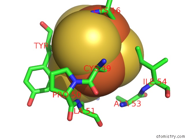
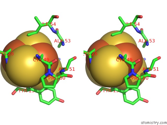
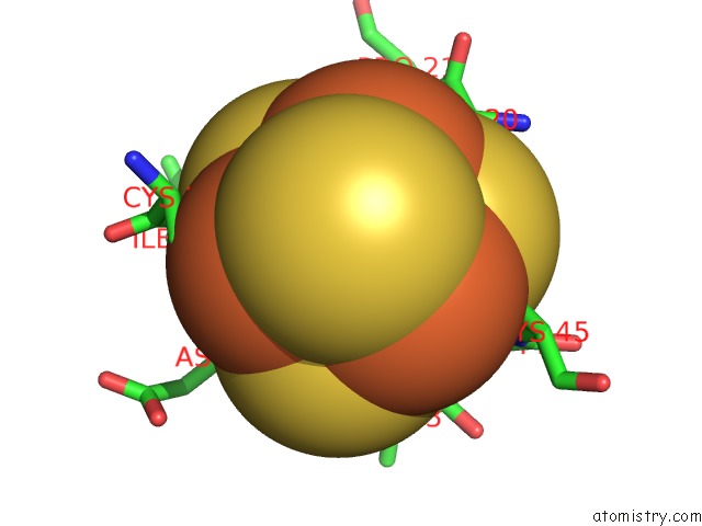
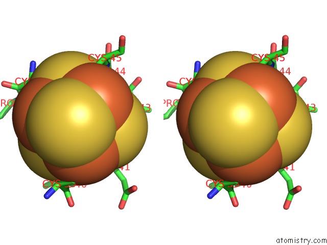
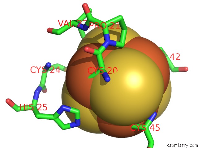
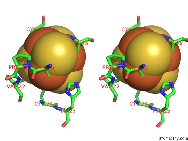
Iron binding site 1 out of 7 in 1f5c
Go back to
Iron binding site 1 out
of 7 in the Crystal Structure of F25H Ferredoxin 1 Mutant From Azotobacter Vinelandii at 1.75 Angstrom Resolution
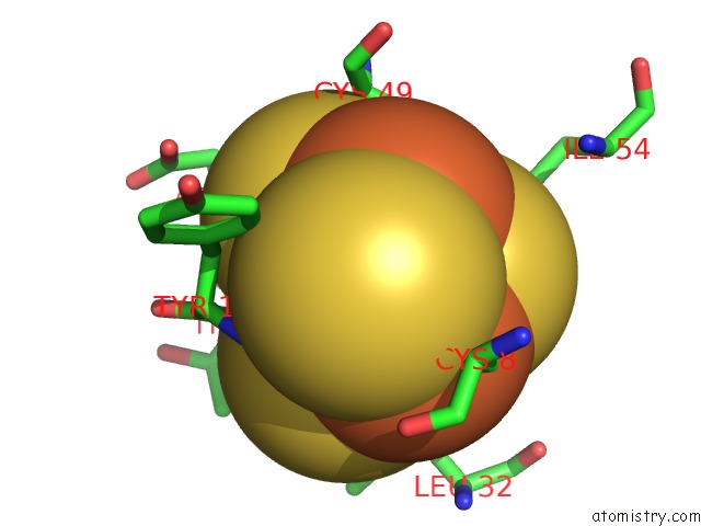
Mono view
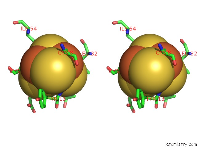
Stereo pair view
Mono view
Stereo pair view
A full contact list of Iron with other atoms in the Fe binding
site number 1 of Crystal Structure of F25H Ferredoxin 1 Mutant From Azotobacter Vinelandii at 1.75 Angstrom Resolution within 5.0Å range:
|
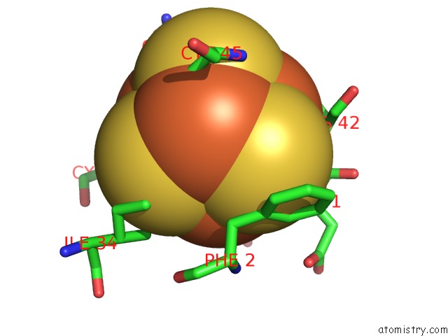
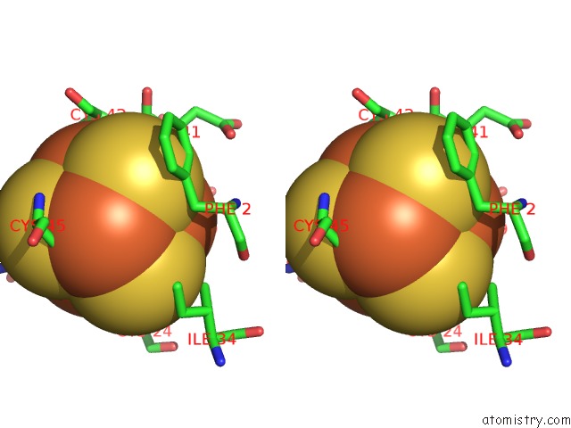
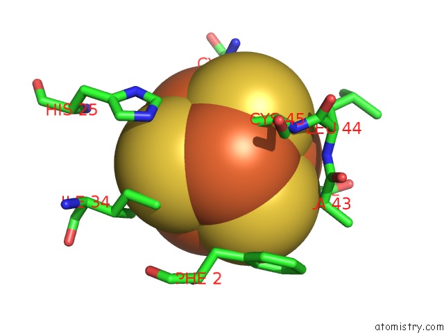
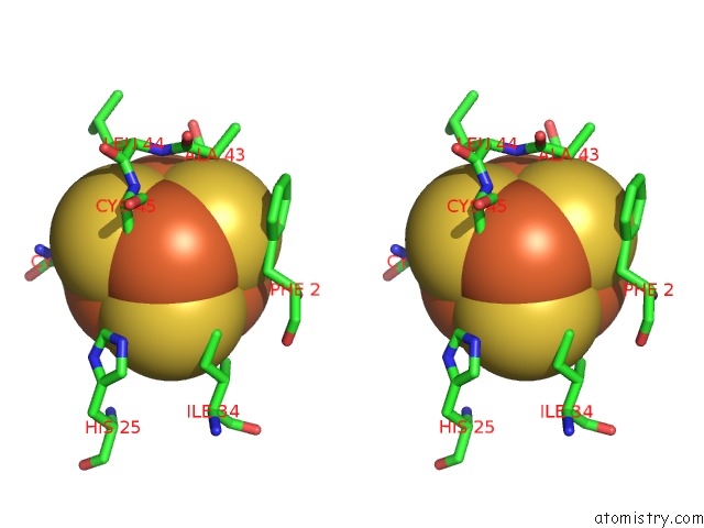
Iron binding site 2 out of 7 in 1f5c
Go back to
Iron binding site 2 out
of 7 in the Crystal Structure of F25H Ferredoxin 1 Mutant From Azotobacter Vinelandii at 1.75 Angstrom Resolution
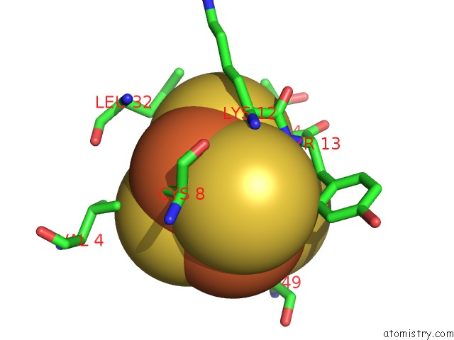
Mono view
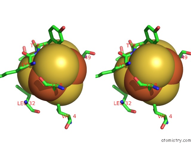
Stereo pair view
Mono view
Stereo pair view
A full contact list of Iron with other atoms in the Fe binding
site number 2 of Crystal Structure of F25H Ferredoxin 1 Mutant From Azotobacter Vinelandii at 1.75 Angstrom Resolution within 5.0Å range:
|
Iron binding site 3 out of 7 in 1f5c
Go back to
Iron binding site 3 out
of 7 in the Crystal Structure of F25H Ferredoxin 1 Mutant From Azotobacter Vinelandii at 1.75 Angstrom Resolution
Mono view
Stereo pair view
Mono view
Stereo pair view
A full contact list of Iron with other atoms in the Fe binding
site number 3 of Crystal Structure of F25H Ferredoxin 1 Mutant From Azotobacter Vinelandii at 1.75 Angstrom Resolution within 5.0Å range:
|
Iron binding site 4 out of 7 in 1f5c
Go back to
Iron binding site 4 out
of 7 in the Crystal Structure of F25H Ferredoxin 1 Mutant From Azotobacter Vinelandii at 1.75 Angstrom Resolution
Mono view
Stereo pair view
Mono view
Stereo pair view
A full contact list of Iron with other atoms in the Fe binding
site number 4 of Crystal Structure of F25H Ferredoxin 1 Mutant From Azotobacter Vinelandii at 1.75 Angstrom Resolution within 5.0Å range:
|
Iron binding site 5 out of 7 in 1f5c
Go back to
Iron binding site 5 out
of 7 in the Crystal Structure of F25H Ferredoxin 1 Mutant From Azotobacter Vinelandii at 1.75 Angstrom Resolution
Mono view
Stereo pair view
Mono view
Stereo pair view
A full contact list of Iron with other atoms in the Fe binding
site number 5 of Crystal Structure of F25H Ferredoxin 1 Mutant From Azotobacter Vinelandii at 1.75 Angstrom Resolution within 5.0Å range:
|
Iron binding site 6 out of 7 in 1f5c
Go back to
Iron binding site 6 out
of 7 in the Crystal Structure of F25H Ferredoxin 1 Mutant From Azotobacter Vinelandii at 1.75 Angstrom Resolution
Mono view
Stereo pair view
Mono view
Stereo pair view
A full contact list of Iron with other atoms in the Fe binding
site number 6 of Crystal Structure of F25H Ferredoxin 1 Mutant From Azotobacter Vinelandii at 1.75 Angstrom Resolution within 5.0Å range:
|
Iron binding site 7 out of 7 in 1f5c
Go back to
Iron binding site 7 out
of 7 in the Crystal Structure of F25H Ferredoxin 1 Mutant From Azotobacter Vinelandii at 1.75 Angstrom Resolution
Mono view
Stereo pair view
Mono view
Stereo pair view
A full contact list of Iron with other atoms in the Fe binding
site number 7 of Crystal Structure of F25H Ferredoxin 1 Mutant From Azotobacter Vinelandii at 1.75 Angstrom Resolution within 5.0Å range:
|
Reference:
K.Chen,
C.A.Bonagura,
G.J.Tilley,
J.P.Mcevoy,
Y.S.Jung,
F.A.Armstrong,
C.D.Stout,
B.K.Burgess.
Crystal Structures of Ferredoxin Variants Exhibiting Large Changes in [Fe-S] Reduction Potential. Nat.Struct.Biol. V. 9 188 2002.
ISSN: ISSN 1072-8368
PubMed: 11875515
Page generated: Sat Aug 3 04:44:05 2024
ISSN: ISSN 1072-8368
PubMed: 11875515
Last articles
Zn in 9J0NZn in 9J0O
Zn in 9J0P
Zn in 9FJX
Zn in 9EKB
Zn in 9C0F
Zn in 9CAH
Zn in 9CH0
Zn in 9CH3
Zn in 9CH1