Iron »
PDB 1etp-1faw »
1f9b »
Iron in PDB 1f9b: Melanin Protein Interaction: X-Ray Structure of the Complex of Mare Lactoferrin with Melanin Monomers
Protein crystallography data
The structure of Melanin Protein Interaction: X-Ray Structure of the Complex of Mare Lactoferrin with Melanin Monomers, PDB code: 1f9b
was solved by
S.Kumar,
T.P.Singh,
A.K.Sharma,
N.Singh,
G.Raman,
with X-Ray Crystallography technique. A brief refinement statistics is given in the table below:
| Resolution Low / High (Å) | 15.00 / 2.70 |
| Space group | P 21 21 21 |
| Cell size a, b, c (Å), α, β, γ (°) | 85.037, 99.815, 103.425, 90.00, 90.00, 90.00 |
| R / Rfree (%) | 21.5 / 28.7 |
Iron Binding Sites:
The binding sites of Iron atom in the Melanin Protein Interaction: X-Ray Structure of the Complex of Mare Lactoferrin with Melanin Monomers
(pdb code 1f9b). This binding sites where shown within
5.0 Angstroms radius around Iron atom.
In total 2 binding sites of Iron where determined in the Melanin Protein Interaction: X-Ray Structure of the Complex of Mare Lactoferrin with Melanin Monomers, PDB code: 1f9b:
Jump to Iron binding site number: 1; 2;
In total 2 binding sites of Iron where determined in the Melanin Protein Interaction: X-Ray Structure of the Complex of Mare Lactoferrin with Melanin Monomers, PDB code: 1f9b:
Jump to Iron binding site number: 1; 2;
Iron binding site 1 out of 2 in 1f9b
Go back to
Iron binding site 1 out
of 2 in the Melanin Protein Interaction: X-Ray Structure of the Complex of Mare Lactoferrin with Melanin Monomers
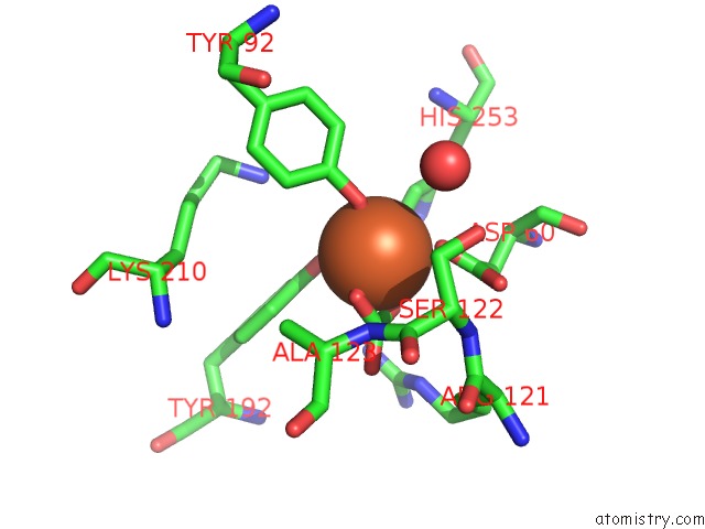
Mono view
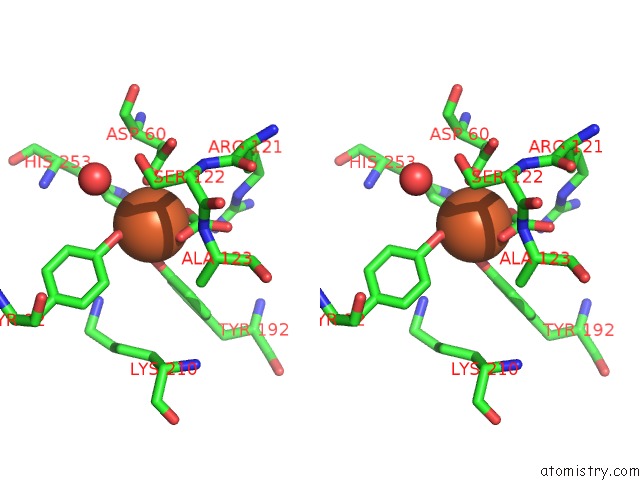
Stereo pair view
Mono view
Stereo pair view
A full contact list of Iron with other atoms in the Fe binding
site number 1 of Melanin Protein Interaction: X-Ray Structure of the Complex of Mare Lactoferrin with Melanin Monomers within 5.0Å range:
|
Iron binding site 2 out of 2 in 1f9b
Go back to
Iron binding site 2 out
of 2 in the Melanin Protein Interaction: X-Ray Structure of the Complex of Mare Lactoferrin with Melanin Monomers
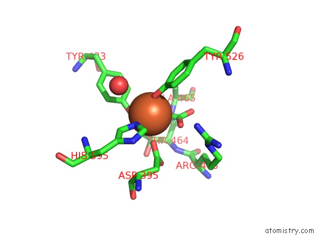
Mono view
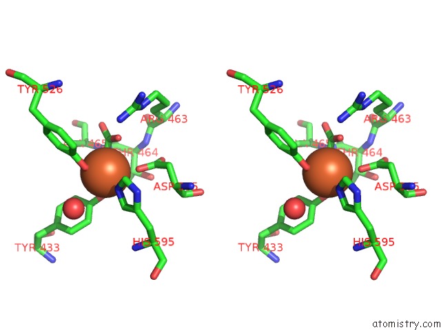
Stereo pair view
Mono view
Stereo pair view
A full contact list of Iron with other atoms in the Fe binding
site number 2 of Melanin Protein Interaction: X-Ray Structure of the Complex of Mare Lactoferrin with Melanin Monomers within 5.0Å range:
|
Reference:
A.K.Sharma,
S.Kumar,
V.Sharma,
A.Nagpal,
N.Singh,
I.Tamboli,
I.Mani,
G.Raman,
T.P.Singh.
Lactoferrin-Melanin Interaction and Its Possible Implications in Melanin Polymerization: Crystal Structure of the Complex Formed Between Mare Lactoferrin and Melanin Monomers at 2.7-A Resolution. Proteins V. 45 229 2001.
ISSN: ISSN 0887-3585
PubMed: 11599026
DOI: 10.1002/PROT.1143
Page generated: Sat Aug 3 04:46:32 2024
ISSN: ISSN 0887-3585
PubMed: 11599026
DOI: 10.1002/PROT.1143
Last articles
Zn in 9MJ5Zn in 9HNW
Zn in 9G0L
Zn in 9FNE
Zn in 9DZN
Zn in 9E0I
Zn in 9D32
Zn in 9DAK
Zn in 8ZXC
Zn in 8ZUF