Iron »
PDB 1etp-1faw »
1fag »
Iron in PDB 1fag: Structure of Cytochrome P450
Enzymatic activity of Structure of Cytochrome P450
All present enzymatic activity of Structure of Cytochrome P450:
1.14.14.1;
1.14.14.1;
Protein crystallography data
The structure of Structure of Cytochrome P450, PDB code: 1fag
was solved by
H.Y.Li,
T.L.Poulos,
with X-Ray Crystallography technique. A brief refinement statistics is given in the table below:
| Resolution Low / High (Å) | 10.00 / 2.70 |
| Space group | C 2 2 21 |
| Cell size a, b, c (Å), α, β, γ (°) | 106.200, 165.200, 223.900, 90.00, 90.00, 90.00 |
| R / Rfree (%) | 24.6 / 35.3 |
Iron Binding Sites:
The binding sites of Iron atom in the Structure of Cytochrome P450
(pdb code 1fag). This binding sites where shown within
5.0 Angstroms radius around Iron atom.
In total 4 binding sites of Iron where determined in the Structure of Cytochrome P450, PDB code: 1fag:
Jump to Iron binding site number: 1; 2; 3; 4;
In total 4 binding sites of Iron where determined in the Structure of Cytochrome P450, PDB code: 1fag:
Jump to Iron binding site number: 1; 2; 3; 4;
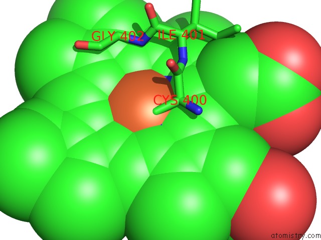
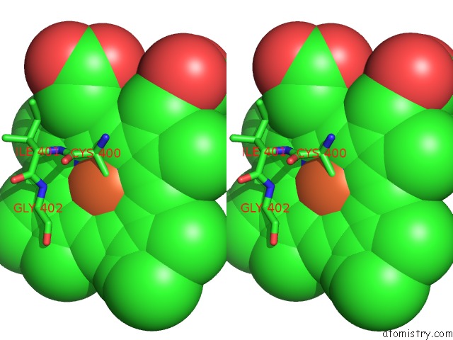
Iron binding site 1 out of 4 in 1fag
Go back to
Iron binding site 1 out
of 4 in the Structure of Cytochrome P450
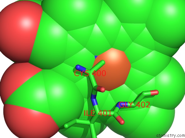
Mono view
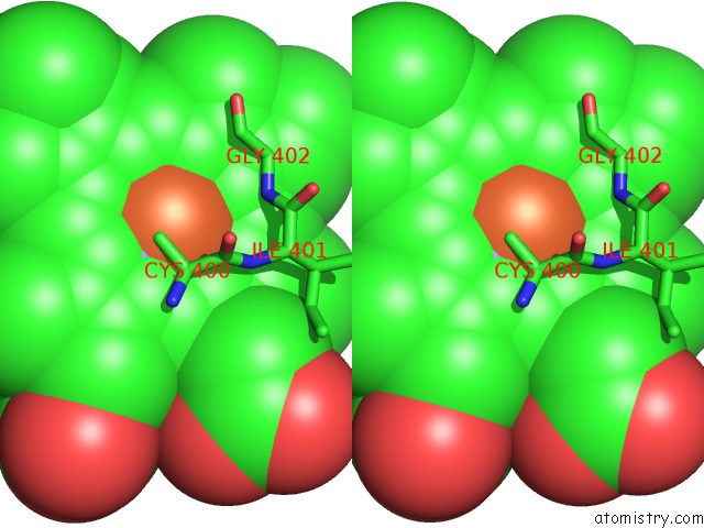
Stereo pair view
Mono view
Stereo pair view
A full contact list of Iron with other atoms in the Fe binding
site number 1 of Structure of Cytochrome P450 within 5.0Å range:
|
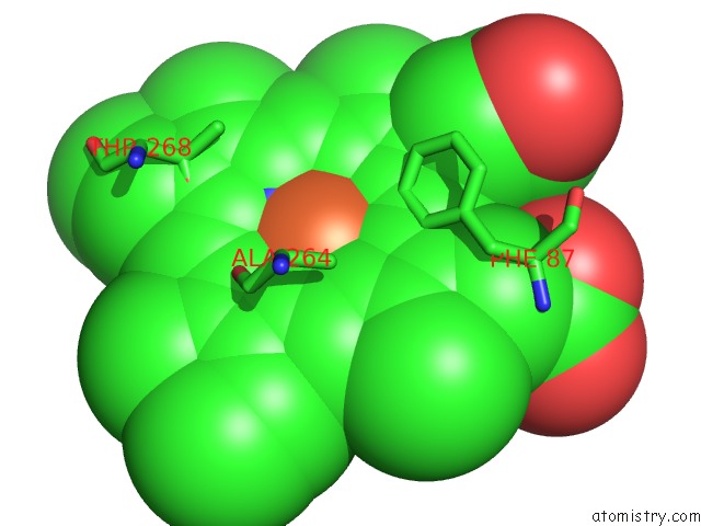
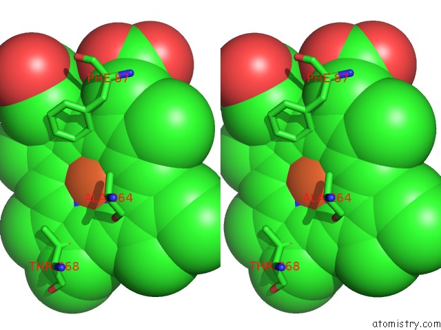
Iron binding site 2 out of 4 in 1fag
Go back to
Iron binding site 2 out
of 4 in the Structure of Cytochrome P450
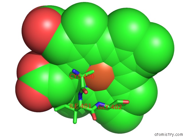
Mono view
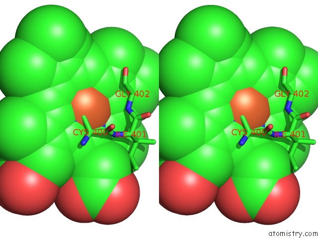
Stereo pair view
Mono view
Stereo pair view
A full contact list of Iron with other atoms in the Fe binding
site number 2 of Structure of Cytochrome P450 within 5.0Å range:
|
Iron binding site 3 out of 4 in 1fag
Go back to
Iron binding site 3 out
of 4 in the Structure of Cytochrome P450
Mono view
Stereo pair view
Mono view
Stereo pair view
A full contact list of Iron with other atoms in the Fe binding
site number 3 of Structure of Cytochrome P450 within 5.0Å range:
|
Iron binding site 4 out of 4 in 1fag
Go back to
Iron binding site 4 out
of 4 in the Structure of Cytochrome P450
Mono view
Stereo pair view
Mono view
Stereo pair view
A full contact list of Iron with other atoms in the Fe binding
site number 4 of Structure of Cytochrome P450 within 5.0Å range:
|
Reference:
H.Li,
T.L.Poulos.
The Structure of the Cytochrome P450BM-3 Haem Domain Complexed with the Fatty Acid Substrate, Palmitoleic Acid. Nat.Struct.Biol. V. 4 140 1997.
ISSN: ISSN 1072-8368
PubMed: 9033595
DOI: 10.1038/NSB0297-140
Page generated: Sat Aug 3 04:46:40 2024
ISSN: ISSN 1072-8368
PubMed: 9033595
DOI: 10.1038/NSB0297-140
Last articles
Zn in 9MJ5Zn in 9HNW
Zn in 9G0L
Zn in 9FNE
Zn in 9DZN
Zn in 9E0I
Zn in 9D32
Zn in 9DAK
Zn in 8ZXC
Zn in 8ZUF