Iron »
PDB 1fca-1fnp »
1ff2 »
Iron in PDB 1ff2: Crystal Structure of the C42D Mutant of Azotobacter Vinelandii 7FE Ferredoxin (Fdi)
Protein crystallography data
The structure of Crystal Structure of the C42D Mutant of Azotobacter Vinelandii 7FE Ferredoxin (Fdi), PDB code: 1ff2
was solved by
C.D.Stout,
B.K.Burgess,
with X-Ray Crystallography technique. A brief refinement statistics is given in the table below:
| Resolution Low / High (Å) | 23.80 / 2.30 |
| Space group | P 41 21 2 |
| Cell size a, b, c (Å), α, β, γ (°) | 55.300, 55.300, 90.590, 90.00, 90.00, 90.00 |
| R / Rfree (%) | 21.7 / 26.9 |
Iron Binding Sites:
The binding sites of Iron atom in the Crystal Structure of the C42D Mutant of Azotobacter Vinelandii 7FE Ferredoxin (Fdi)
(pdb code 1ff2). This binding sites where shown within
5.0 Angstroms radius around Iron atom.
In total 7 binding sites of Iron where determined in the Crystal Structure of the C42D Mutant of Azotobacter Vinelandii 7FE Ferredoxin (Fdi), PDB code: 1ff2:
Jump to Iron binding site number: 1; 2; 3; 4; 5; 6; 7;
In total 7 binding sites of Iron where determined in the Crystal Structure of the C42D Mutant of Azotobacter Vinelandii 7FE Ferredoxin (Fdi), PDB code: 1ff2:
Jump to Iron binding site number: 1; 2; 3; 4; 5; 6; 7;
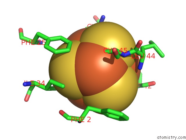
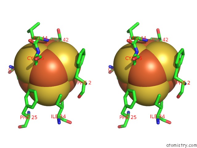
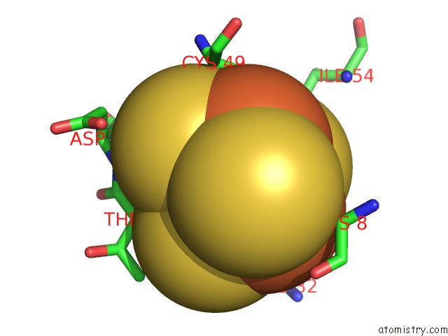
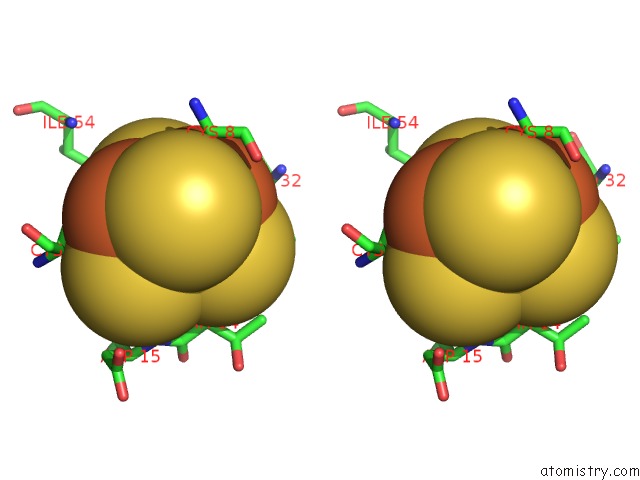
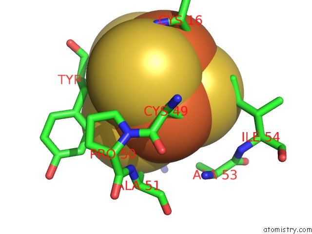
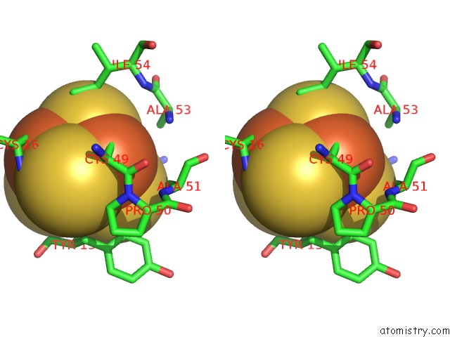
Iron binding site 1 out of 7 in 1ff2
Go back to
Iron binding site 1 out
of 7 in the Crystal Structure of the C42D Mutant of Azotobacter Vinelandii 7FE Ferredoxin (Fdi)
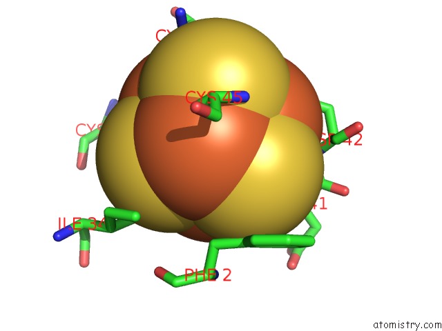
Mono view
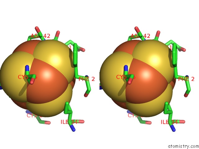
Stereo pair view
Mono view
Stereo pair view
A full contact list of Iron with other atoms in the Fe binding
site number 1 of Crystal Structure of the C42D Mutant of Azotobacter Vinelandii 7FE Ferredoxin (Fdi) within 5.0Å range:
|
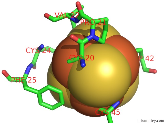
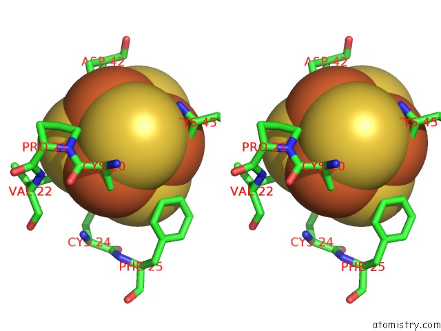
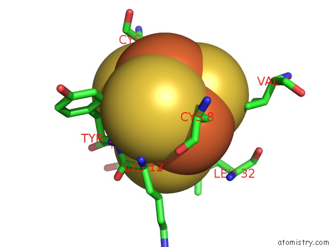
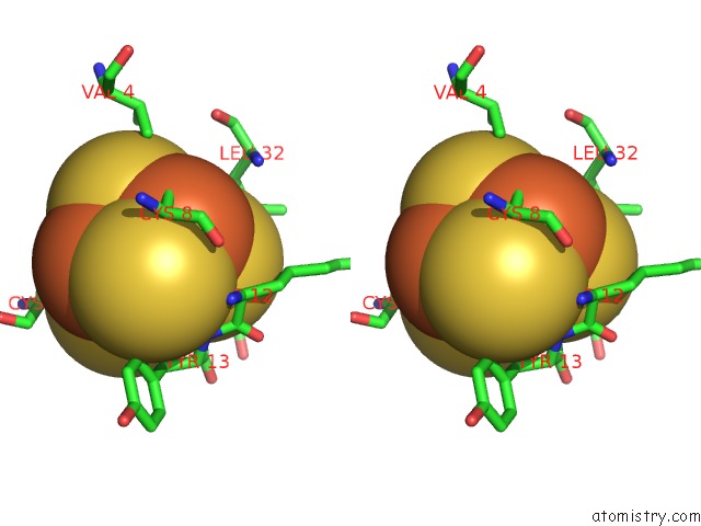
Iron binding site 2 out of 7 in 1ff2
Go back to
Iron binding site 2 out
of 7 in the Crystal Structure of the C42D Mutant of Azotobacter Vinelandii 7FE Ferredoxin (Fdi)
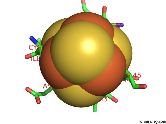
Mono view
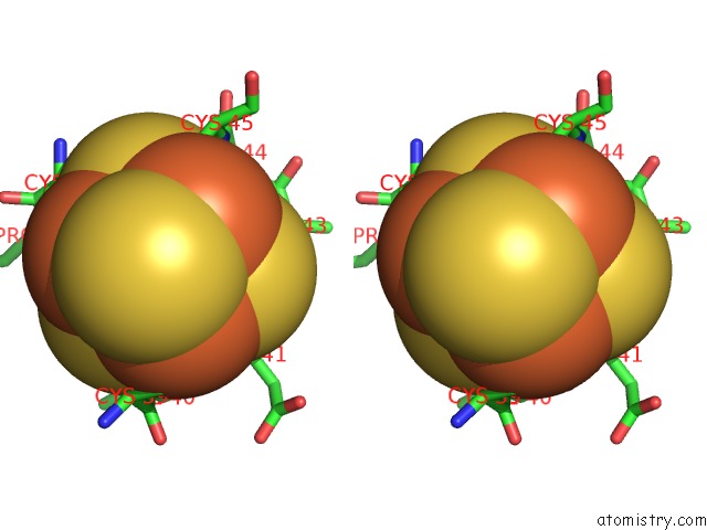
Stereo pair view
Mono view
Stereo pair view
A full contact list of Iron with other atoms in the Fe binding
site number 2 of Crystal Structure of the C42D Mutant of Azotobacter Vinelandii 7FE Ferredoxin (Fdi) within 5.0Å range:
|
Iron binding site 3 out of 7 in 1ff2
Go back to
Iron binding site 3 out
of 7 in the Crystal Structure of the C42D Mutant of Azotobacter Vinelandii 7FE Ferredoxin (Fdi)
Mono view
Stereo pair view
Mono view
Stereo pair view
A full contact list of Iron with other atoms in the Fe binding
site number 3 of Crystal Structure of the C42D Mutant of Azotobacter Vinelandii 7FE Ferredoxin (Fdi) within 5.0Å range:
|
Iron binding site 4 out of 7 in 1ff2
Go back to
Iron binding site 4 out
of 7 in the Crystal Structure of the C42D Mutant of Azotobacter Vinelandii 7FE Ferredoxin (Fdi)
Mono view
Stereo pair view
Mono view
Stereo pair view
A full contact list of Iron with other atoms in the Fe binding
site number 4 of Crystal Structure of the C42D Mutant of Azotobacter Vinelandii 7FE Ferredoxin (Fdi) within 5.0Å range:
|
Iron binding site 5 out of 7 in 1ff2
Go back to
Iron binding site 5 out
of 7 in the Crystal Structure of the C42D Mutant of Azotobacter Vinelandii 7FE Ferredoxin (Fdi)
Mono view
Stereo pair view
Mono view
Stereo pair view
A full contact list of Iron with other atoms in the Fe binding
site number 5 of Crystal Structure of the C42D Mutant of Azotobacter Vinelandii 7FE Ferredoxin (Fdi) within 5.0Å range:
|
Iron binding site 6 out of 7 in 1ff2
Go back to
Iron binding site 6 out
of 7 in the Crystal Structure of the C42D Mutant of Azotobacter Vinelandii 7FE Ferredoxin (Fdi)
Mono view
Stereo pair view
Mono view
Stereo pair view
A full contact list of Iron with other atoms in the Fe binding
site number 6 of Crystal Structure of the C42D Mutant of Azotobacter Vinelandii 7FE Ferredoxin (Fdi) within 5.0Å range:
|
Iron binding site 7 out of 7 in 1ff2
Go back to
Iron binding site 7 out
of 7 in the Crystal Structure of the C42D Mutant of Azotobacter Vinelandii 7FE Ferredoxin (Fdi)
Mono view
Stereo pair view
Mono view
Stereo pair view
A full contact list of Iron with other atoms in the Fe binding
site number 7 of Crystal Structure of the C42D Mutant of Azotobacter Vinelandii 7FE Ferredoxin (Fdi) within 5.0Å range:
|
Reference:
Y.S.Jung,
C.A.Bonagura,
G.J.Tilley,
H.S.Gao-Sheridan,
F.A.Armstrong,
C.D.Stout,
B.K.Burgess.
Structure of C42D Azotobacter Vinelandii Fdi. A Cys-X-X-Asp-X-X-Cys Motif Ligates An Air-Stable [4FE-4S]2+/+ Cluster J.Biol.Chem. V. 275 36974 2000.
ISSN: ISSN 0021-9258
PubMed: 10961993
DOI: 10.1074/JBC.M004947200
Page generated: Sat Aug 3 04:56:43 2024
ISSN: ISSN 0021-9258
PubMed: 10961993
DOI: 10.1074/JBC.M004947200
Last articles
F in 4LWVF in 4LW1
F in 4LWU
F in 4LTS
F in 4LUD
F in 4LUV
F in 4LOQ
F in 4LQG
F in 4LPB
F in 4LOP