Iron »
PDB 1fca-1fnp »
1fiq »
Iron in PDB 1fiq: Crystal Structure of Xanthine Oxidase From Bovine Milk
Enzymatic activity of Crystal Structure of Xanthine Oxidase From Bovine Milk
All present enzymatic activity of Crystal Structure of Xanthine Oxidase From Bovine Milk:
1.1.3.22;
1.1.3.22;
Protein crystallography data
The structure of Crystal Structure of Xanthine Oxidase From Bovine Milk, PDB code: 1fiq
was solved by
C.Enroth,
B.T.Eger,
K.Okamoto,
T.Nishino,
T.Nishino,
E.F.Pai,
with X-Ray Crystallography technique. A brief refinement statistics is given in the table below:
| Resolution Low / High (Å) | 8.00 / 2.50 |
| Space group | C 2 2 21 |
| Cell size a, b, c (Å), α, β, γ (°) | 117.831, 165.403, 154.447, 90.00, 90.00, 90.00 |
| R / Rfree (%) | 21.2 / 27.5 |
Other elements in 1fiq:
The structure of Crystal Structure of Xanthine Oxidase From Bovine Milk also contains other interesting chemical elements:
| Molybdenum | (Mo) | 1 atom |
Iron Binding Sites:
The binding sites of Iron atom in the Crystal Structure of Xanthine Oxidase From Bovine Milk
(pdb code 1fiq). This binding sites where shown within
5.0 Angstroms radius around Iron atom.
In total 4 binding sites of Iron where determined in the Crystal Structure of Xanthine Oxidase From Bovine Milk, PDB code: 1fiq:
Jump to Iron binding site number: 1; 2; 3; 4;
In total 4 binding sites of Iron where determined in the Crystal Structure of Xanthine Oxidase From Bovine Milk, PDB code: 1fiq:
Jump to Iron binding site number: 1; 2; 3; 4;
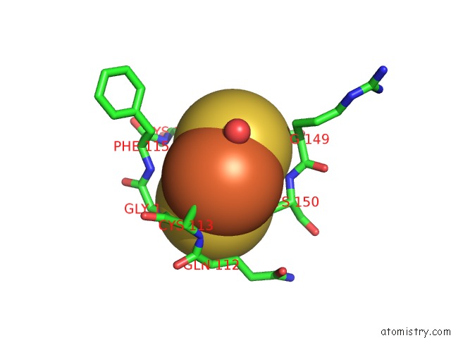
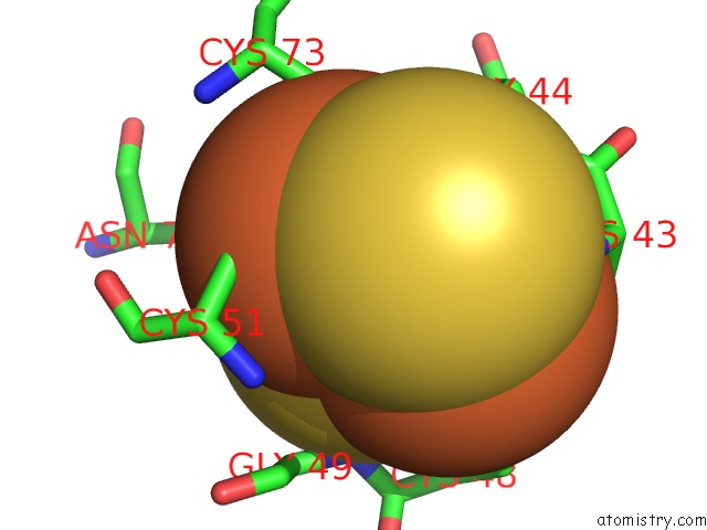
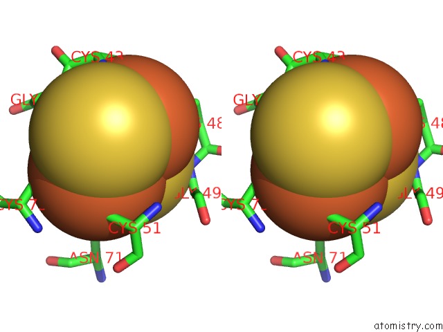
Iron binding site 1 out of 4 in 1fiq
Go back to
Iron binding site 1 out
of 4 in the Crystal Structure of Xanthine Oxidase From Bovine Milk
Mono view
Stereo pair view
Mono view
Stereo pair view
A full contact list of Iron with other atoms in the Fe binding
site number 1 of Crystal Structure of Xanthine Oxidase From Bovine Milk within 5.0Å range:
|
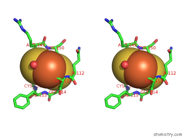
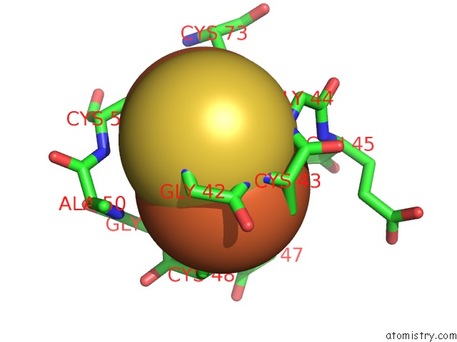
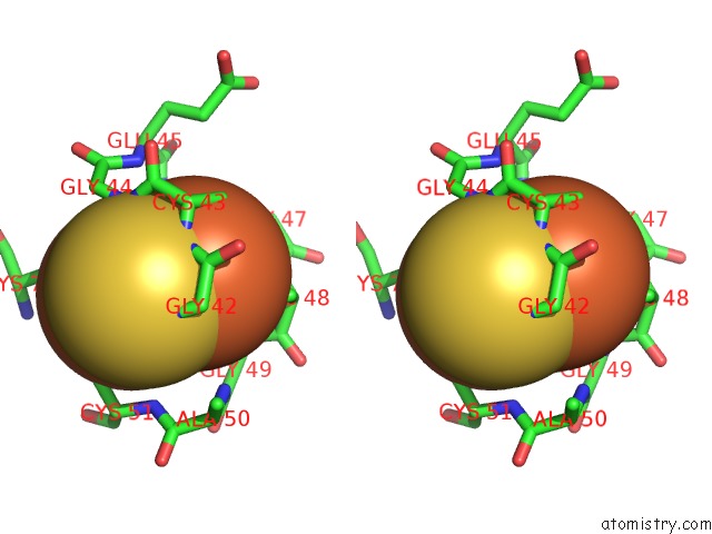
Iron binding site 2 out of 4 in 1fiq
Go back to
Iron binding site 2 out
of 4 in the Crystal Structure of Xanthine Oxidase From Bovine Milk
Mono view
Stereo pair view
Mono view
Stereo pair view
A full contact list of Iron with other atoms in the Fe binding
site number 2 of Crystal Structure of Xanthine Oxidase From Bovine Milk within 5.0Å range:
|
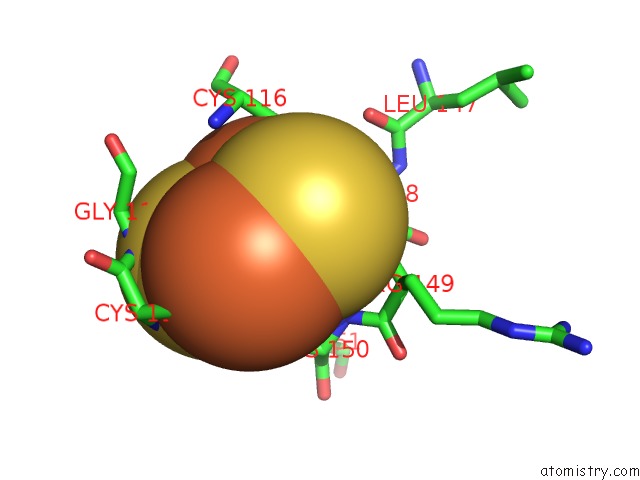
Iron binding site 3 out of 4 in 1fiq
Go back to
Iron binding site 3 out
of 4 in the Crystal Structure of Xanthine Oxidase From Bovine Milk
Mono view
Stereo pair view
Mono view
Stereo pair view
A full contact list of Iron with other atoms in the Fe binding
site number 3 of Crystal Structure of Xanthine Oxidase From Bovine Milk within 5.0Å range:
|
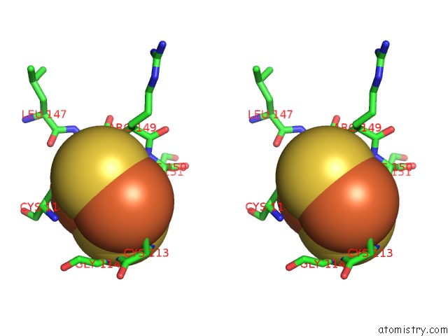
Iron binding site 4 out of 4 in 1fiq
Go back to
Iron binding site 4 out
of 4 in the Crystal Structure of Xanthine Oxidase From Bovine Milk
Mono view
Stereo pair view
Mono view
Stereo pair view
A full contact list of Iron with other atoms in the Fe binding
site number 4 of Crystal Structure of Xanthine Oxidase From Bovine Milk within 5.0Å range:
|
Reference:
C.Enroth,
B.T.Eger,
K.Okamoto,
T.Nishino,
T.Nishino,
E.F.Pai.
Crystal Structures of Bovine Milk Xanthine Dehydrogenase and Xanthine Oxidase: Structure-Based Mechanism of Conversion. Proc.Natl.Acad.Sci.Usa V. 97 10723 2000.
ISSN: ISSN 0027-8424
PubMed: 11005854
DOI: 10.1073/PNAS.97.20.10723
Page generated: Sat Aug 3 05:06:26 2024
ISSN: ISSN 0027-8424
PubMed: 11005854
DOI: 10.1073/PNAS.97.20.10723
Last articles
Zn in 9MJ5Zn in 9HNW
Zn in 9G0L
Zn in 9FNE
Zn in 9DZN
Zn in 9E0I
Zn in 9D32
Zn in 9DAK
Zn in 8ZXC
Zn in 8ZUF