Iron »
PDB 1fca-1fnp »
1flp »
Iron in PDB 1flp: Structure of the Sulfide-Reactive Hemoglobin From the Clam Lucina Pectinata: Crystallographic Analysis at 1.5 Angstroms Resolution
Protein crystallography data
The structure of Structure of the Sulfide-Reactive Hemoglobin From the Clam Lucina Pectinata: Crystallographic Analysis at 1.5 Angstroms Resolution, PDB code: 1flp
was solved by
M.Rizzi,
J.B.Wittenberg,
P.Ascenzi,
M.Fasano,
A.Coda,
M.Bolognesi,
with X-Ray Crystallography technique. A brief refinement statistics is given in the table below:
| Resolution Low / High (Å) | 15.00 / 1.50 |
| Space group | P 1 21 1 |
| Cell size a, b, c (Å), α, β, γ (°) | 37.970, 38.390, 42.650, 90.00, 97.40, 90.00 |
| R / Rfree (%) | n/a / n/a |
Iron Binding Sites:
The binding sites of Iron atom in the Structure of the Sulfide-Reactive Hemoglobin From the Clam Lucina Pectinata: Crystallographic Analysis at 1.5 Angstroms Resolution
(pdb code 1flp). This binding sites where shown within
5.0 Angstroms radius around Iron atom.
In total only one binding site of Iron was determined in the Structure of the Sulfide-Reactive Hemoglobin From the Clam Lucina Pectinata: Crystallographic Analysis at 1.5 Angstroms Resolution, PDB code: 1flp:
In total only one binding site of Iron was determined in the Structure of the Sulfide-Reactive Hemoglobin From the Clam Lucina Pectinata: Crystallographic Analysis at 1.5 Angstroms Resolution, PDB code: 1flp:
Iron binding site 1 out of 1 in 1flp
Go back to
Iron binding site 1 out
of 1 in the Structure of the Sulfide-Reactive Hemoglobin From the Clam Lucina Pectinata: Crystallographic Analysis at 1.5 Angstroms Resolution
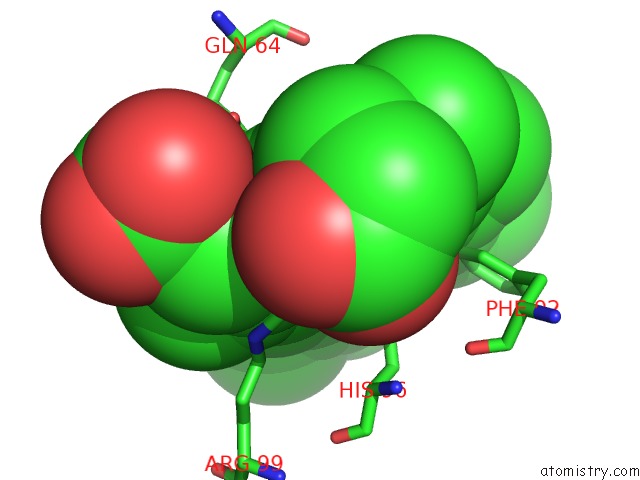
Mono view
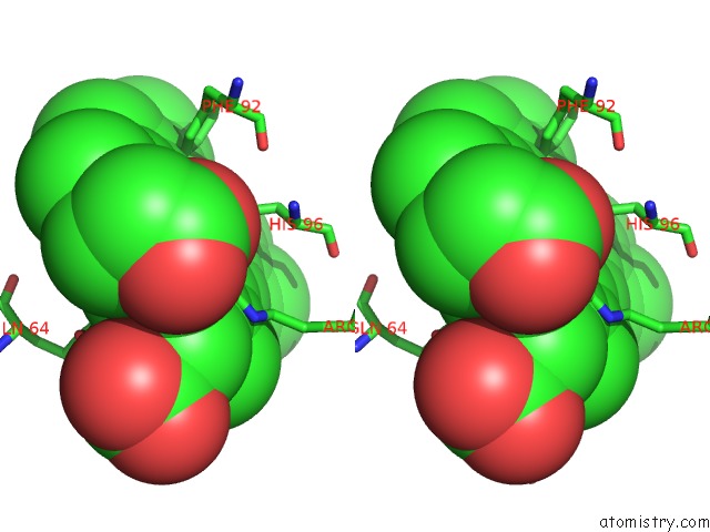
Stereo pair view
Mono view
Stereo pair view
A full contact list of Iron with other atoms in the Fe binding
site number 1 of Structure of the Sulfide-Reactive Hemoglobin From the Clam Lucina Pectinata: Crystallographic Analysis at 1.5 Angstroms Resolution within 5.0Å range:
|
Reference:
M.Rizzi,
J.B.Wittenberg,
A.Coda,
M.Fasano,
P.Ascenzi,
M.Bolognesi.
Structure of the Sulfide-Reactive Hemoglobin From the Clam Lucina Pectinata. Crystallographic Analysis at 1.5 A Resolution. J.Mol.Biol. V. 244 86 1994.
ISSN: ISSN 0022-2836
PubMed: 7966324
DOI: 10.1006/JMBI.1994.1706
Page generated: Sat Aug 3 05:07:04 2024
ISSN: ISSN 0022-2836
PubMed: 7966324
DOI: 10.1006/JMBI.1994.1706
Last articles
Zn in 9MJ5Zn in 9HNW
Zn in 9G0L
Zn in 9FNE
Zn in 9DZN
Zn in 9E0I
Zn in 9D32
Zn in 9DAK
Zn in 8ZXC
Zn in 8ZUF