Iron »
PDB 1fz2-1gek »
1fz4 »
Iron in PDB 1fz4: Methane Monooxygenase Hydroxylase, Form III Soaked at pH 8.5 (0.1 M Tris)
Enzymatic activity of Methane Monooxygenase Hydroxylase, Form III Soaked at pH 8.5 (0.1 M Tris)
All present enzymatic activity of Methane Monooxygenase Hydroxylase, Form III Soaked at pH 8.5 (0.1 M Tris):
1.14.13.25;
1.14.13.25;
Protein crystallography data
The structure of Methane Monooxygenase Hydroxylase, Form III Soaked at pH 8.5 (0.1 M Tris), PDB code: 1fz4
was solved by
D.A.Whittington,
S.J.Lippard,
with X-Ray Crystallography technique. A brief refinement statistics is given in the table below:
| Resolution Low / High (Å) | 29.70 / 2.38 |
| Space group | P 21 21 21 |
| Cell size a, b, c (Å), α, β, γ (°) | 70.890, 171.520, 221.690, 90.00, 90.00, 90.00 |
| R / Rfree (%) | 22.3 / 25.6 |
Other elements in 1fz4:
The structure of Methane Monooxygenase Hydroxylase, Form III Soaked at pH 8.5 (0.1 M Tris) also contains other interesting chemical elements:
| Calcium | (Ca) | 3 atoms |
Iron Binding Sites:
The binding sites of Iron atom in the Methane Monooxygenase Hydroxylase, Form III Soaked at pH 8.5 (0.1 M Tris)
(pdb code 1fz4). This binding sites where shown within
5.0 Angstroms radius around Iron atom.
In total 4 binding sites of Iron where determined in the Methane Monooxygenase Hydroxylase, Form III Soaked at pH 8.5 (0.1 M Tris), PDB code: 1fz4:
Jump to Iron binding site number: 1; 2; 3; 4;
In total 4 binding sites of Iron where determined in the Methane Monooxygenase Hydroxylase, Form III Soaked at pH 8.5 (0.1 M Tris), PDB code: 1fz4:
Jump to Iron binding site number: 1; 2; 3; 4;
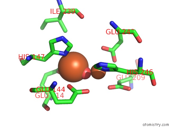
Iron binding site 1 out of 4 in 1fz4
Go back to
Iron binding site 1 out
of 4 in the Methane Monooxygenase Hydroxylase, Form III Soaked at pH 8.5 (0.1 M Tris)
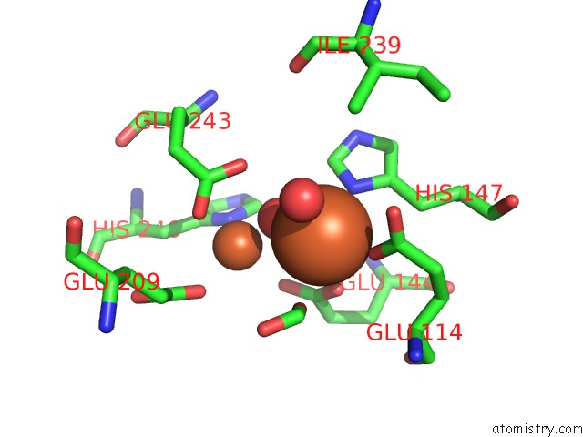
Mono view
Stereo pair view
Mono view
Stereo pair view
A full contact list of Iron with other atoms in the Fe binding
site number 1 of Methane Monooxygenase Hydroxylase, Form III Soaked at pH 8.5 (0.1 M Tris) within 5.0Å range:
|
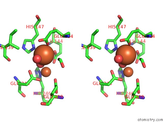
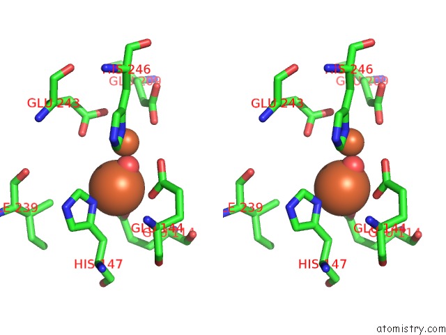
Iron binding site 2 out of 4 in 1fz4
Go back to
Iron binding site 2 out
of 4 in the Methane Monooxygenase Hydroxylase, Form III Soaked at pH 8.5 (0.1 M Tris)
Mono view
Stereo pair view
Mono view
Stereo pair view
A full contact list of Iron with other atoms in the Fe binding
site number 2 of Methane Monooxygenase Hydroxylase, Form III Soaked at pH 8.5 (0.1 M Tris) within 5.0Å range:
|
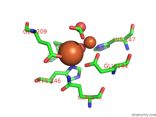
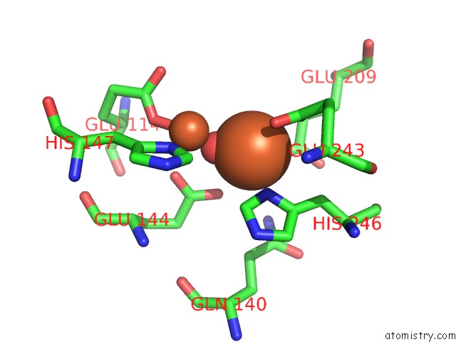
Iron binding site 3 out of 4 in 1fz4
Go back to
Iron binding site 3 out
of 4 in the Methane Monooxygenase Hydroxylase, Form III Soaked at pH 8.5 (0.1 M Tris)
Mono view
Stereo pair view
Mono view
Stereo pair view
A full contact list of Iron with other atoms in the Fe binding
site number 3 of Methane Monooxygenase Hydroxylase, Form III Soaked at pH 8.5 (0.1 M Tris) within 5.0Å range:
|
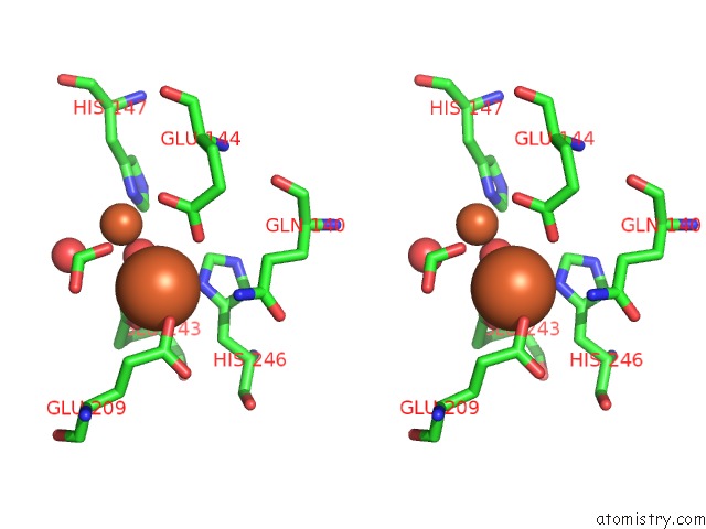
Iron binding site 4 out of 4 in 1fz4
Go back to
Iron binding site 4 out
of 4 in the Methane Monooxygenase Hydroxylase, Form III Soaked at pH 8.5 (0.1 M Tris)
Mono view
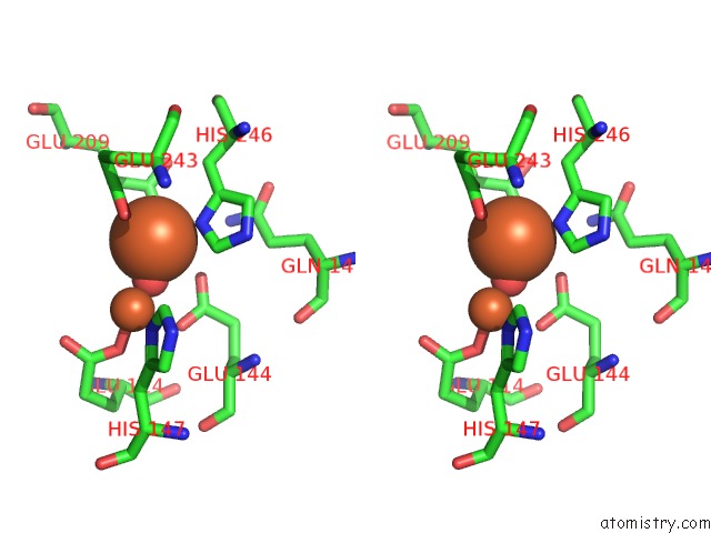
Stereo pair view
Mono view
Stereo pair view
A full contact list of Iron with other atoms in the Fe binding
site number 4 of Methane Monooxygenase Hydroxylase, Form III Soaked at pH 8.5 (0.1 M Tris) within 5.0Å range:
|
Reference:
D.A.Whittington,
S.J.Lippard.
Crystal Structures of the Soluble Methane Monooxygenase Hydroxylase From Methylococcus Capsulatus (Bath) Demonstrating Geometrical Variability at the Dinuclear Iron Active Site. J.Am.Chem.Soc. V. 123 827 2001.
ISSN: ISSN 0002-7863
PubMed: 11456616
DOI: 10.1021/JA003240N
Page generated: Wed Jul 16 14:36:46 2025
ISSN: ISSN 0002-7863
PubMed: 11456616
DOI: 10.1021/JA003240N
Last articles
Fe in 2YXOFe in 2YRS
Fe in 2YXC
Fe in 2YNM
Fe in 2YVJ
Fe in 2YP1
Fe in 2YU2
Fe in 2YU1
Fe in 2YQB
Fe in 2YOO