Iron »
PDB 1fz2-1gek »
1g6b »
Iron in PDB 1g6b: Crystal Structure of P47S Mutant of Ferredoxin I
Protein crystallography data
The structure of Crystal Structure of P47S Mutant of Ferredoxin I, PDB code: 1g6b
was solved by
C.D.Stout,
B.K.Burgess,
C.A.Bonagura,
Y.S.Jung,
with X-Ray Crystallography technique. A brief refinement statistics is given in the table below:
| Resolution Low / High (Å) | 24.00 / 1.90 |
| Space group | P 41 21 2 |
| Cell size a, b, c (Å), α, β, γ (°) | 54.970, 54.970, 92.110, 90.00, 90.00, 90.00 |
| R / Rfree (%) | 24 / 26.6 |
Iron Binding Sites:
The binding sites of Iron atom in the Crystal Structure of P47S Mutant of Ferredoxin I
(pdb code 1g6b). This binding sites where shown within
5.0 Angstroms radius around Iron atom.
In total 7 binding sites of Iron where determined in the Crystal Structure of P47S Mutant of Ferredoxin I, PDB code: 1g6b:
Jump to Iron binding site number: 1; 2; 3; 4; 5; 6; 7;
In total 7 binding sites of Iron where determined in the Crystal Structure of P47S Mutant of Ferredoxin I, PDB code: 1g6b:
Jump to Iron binding site number: 1; 2; 3; 4; 5; 6; 7;
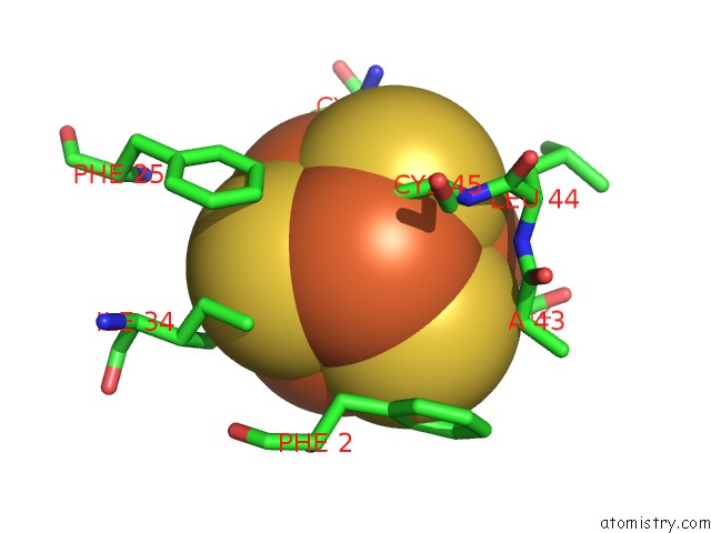
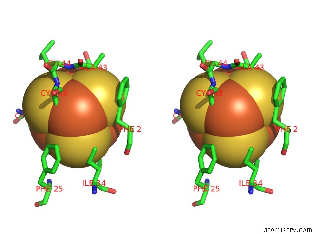
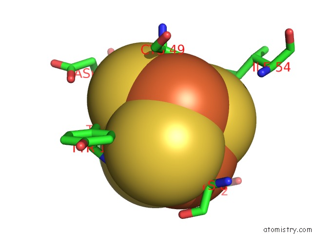
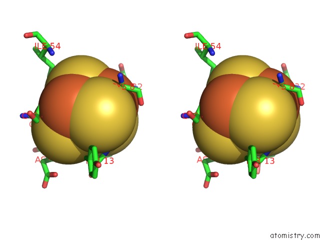
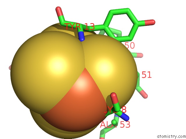
Iron binding site 1 out of 7 in 1g6b
Go back to
Iron binding site 1 out
of 7 in the Crystal Structure of P47S Mutant of Ferredoxin I
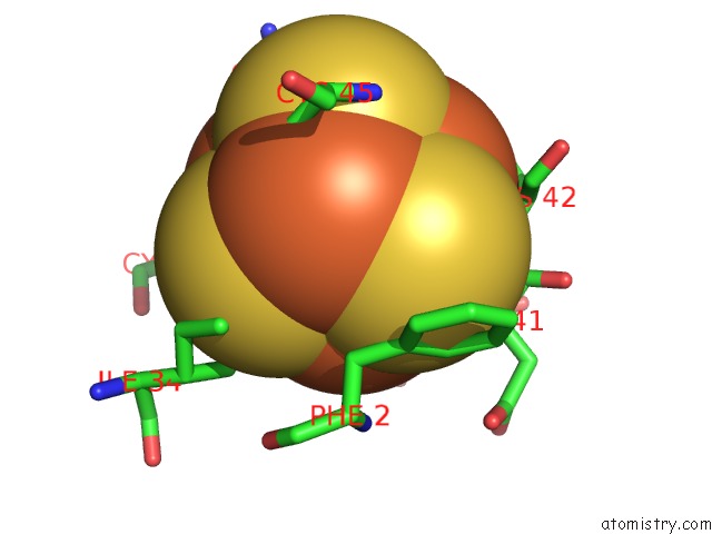
Mono view
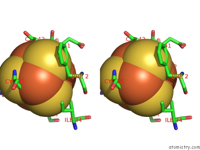
Stereo pair view
Mono view
Stereo pair view
A full contact list of Iron with other atoms in the Fe binding
site number 1 of Crystal Structure of P47S Mutant of Ferredoxin I within 5.0Å range:
|
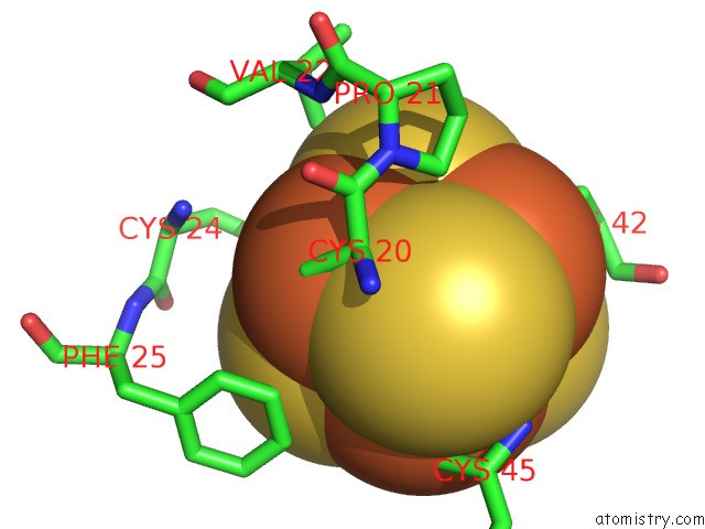
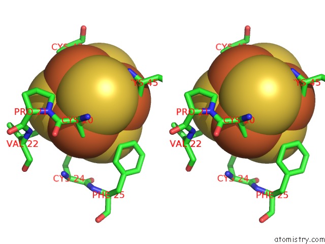
Iron binding site 2 out of 7 in 1g6b
Go back to
Iron binding site 2 out
of 7 in the Crystal Structure of P47S Mutant of Ferredoxin I
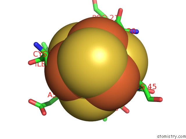
Mono view
Stereo pair view
Mono view
Stereo pair view
A full contact list of Iron with other atoms in the Fe binding
site number 2 of Crystal Structure of P47S Mutant of Ferredoxin I within 5.0Å range:
|
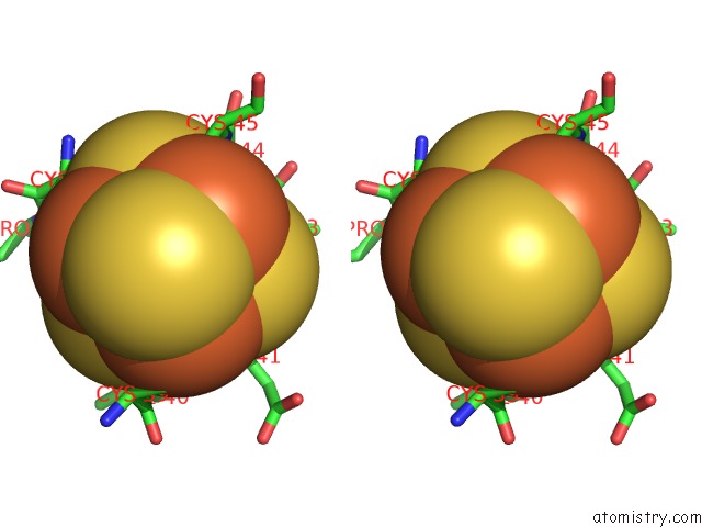
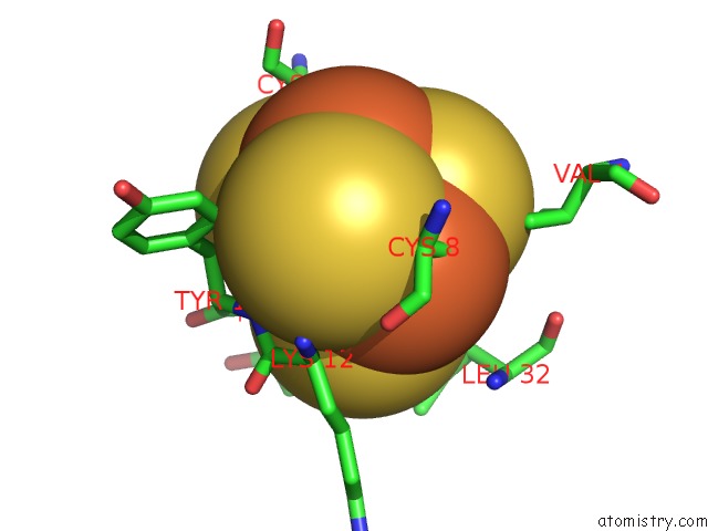
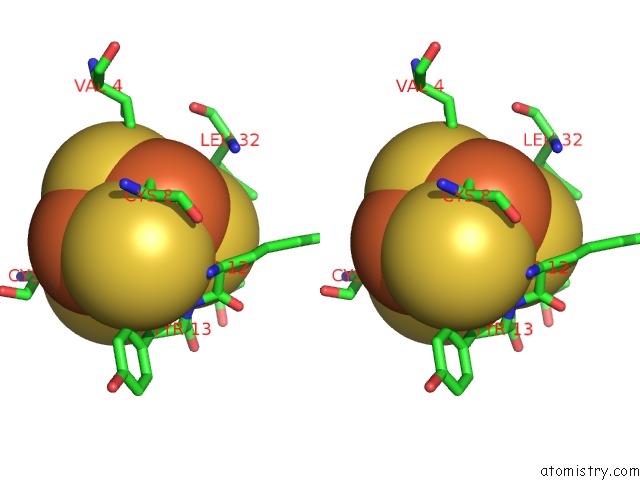
Iron binding site 3 out of 7 in 1g6b
Go back to
Iron binding site 3 out
of 7 in the Crystal Structure of P47S Mutant of Ferredoxin I
Mono view
Stereo pair view
Mono view
Stereo pair view
A full contact list of Iron with other atoms in the Fe binding
site number 3 of Crystal Structure of P47S Mutant of Ferredoxin I within 5.0Å range:
|
Iron binding site 4 out of 7 in 1g6b
Go back to
Iron binding site 4 out
of 7 in the Crystal Structure of P47S Mutant of Ferredoxin I
Mono view
Stereo pair view
Mono view
Stereo pair view
A full contact list of Iron with other atoms in the Fe binding
site number 4 of Crystal Structure of P47S Mutant of Ferredoxin I within 5.0Å range:
|
Iron binding site 5 out of 7 in 1g6b
Go back to
Iron binding site 5 out
of 7 in the Crystal Structure of P47S Mutant of Ferredoxin I
Mono view
Stereo pair view
Mono view
Stereo pair view
A full contact list of Iron with other atoms in the Fe binding
site number 5 of Crystal Structure of P47S Mutant of Ferredoxin I within 5.0Å range:
|
Iron binding site 6 out of 7 in 1g6b
Go back to
Iron binding site 6 out
of 7 in the Crystal Structure of P47S Mutant of Ferredoxin I
Mono view
Stereo pair view
Mono view
Stereo pair view
A full contact list of Iron with other atoms in the Fe binding
site number 6 of Crystal Structure of P47S Mutant of Ferredoxin I within 5.0Å range:
|
Iron binding site 7 out of 7 in 1g6b
Go back to
Iron binding site 7 out
of 7 in the Crystal Structure of P47S Mutant of Ferredoxin I
Mono view

Stereo pair view
Mono view
Stereo pair view
A full contact list of Iron with other atoms in the Fe binding
site number 7 of Crystal Structure of P47S Mutant of Ferredoxin I within 5.0Å range:
|
Reference:
K.Chen,
Y.S.Jung,
C.A.Bonagura,
G.J.Tilley,
G.S.Prasad,
V.Sridhar,
F.A.Armstrong,
C.D.Stout,
B.K.Burgess.
Azotobacter Vinelandii Ferredoxin I: A Sequence and Structure Comparison Approach to Alteration of [4FE-4S]2+/+ Reduction Potential. J.Biol.Chem. V. 277 5603 2002.
ISSN: ISSN 0021-9258
PubMed: 11704670
DOI: 10.1074/JBC.M108916200
Page generated: Sat Aug 3 05:45:36 2024
ISSN: ISSN 0021-9258
PubMed: 11704670
DOI: 10.1074/JBC.M108916200
Last articles
F in 7PK3F in 7PJC
F in 7PJ2
F in 7PHN
F in 7PG6
F in 7PHJ
F in 7PAV
F in 7PH1
F in 7PCD
F in 7P80