Iron »
PDB 1fz2-1gek »
1gbv »
Iron in PDB 1gbv: (Alpha-Oxy, Beta-(C112G)Deoxy) T-State Human Hemoglobin
Protein crystallography data
The structure of (Alpha-Oxy, Beta-(C112G)Deoxy) T-State Human Hemoglobin, PDB code: 1gbv
was solved by
G.B.Vasquez,
X.Ji,
I.Pechik,
C.Fronticelli,
G.L.Gilliland,
with X-Ray Crystallography technique. A brief refinement statistics is given in the table below:
| Resolution Low / High (Å) | 6.00 / 2.00 |
| Space group | P 1 21 1 |
| Cell size a, b, c (Å), α, β, γ (°) | 63.180, 83.560, 53.730, 90.00, 99.60, 90.00 |
| R / Rfree (%) | n/a / n/a |
Iron Binding Sites:
The binding sites of Iron atom in the (Alpha-Oxy, Beta-(C112G)Deoxy) T-State Human Hemoglobin
(pdb code 1gbv). This binding sites where shown within
5.0 Angstroms radius around Iron atom.
In total 4 binding sites of Iron where determined in the (Alpha-Oxy, Beta-(C112G)Deoxy) T-State Human Hemoglobin, PDB code: 1gbv:
Jump to Iron binding site number: 1; 2; 3; 4;
In total 4 binding sites of Iron where determined in the (Alpha-Oxy, Beta-(C112G)Deoxy) T-State Human Hemoglobin, PDB code: 1gbv:
Jump to Iron binding site number: 1; 2; 3; 4;
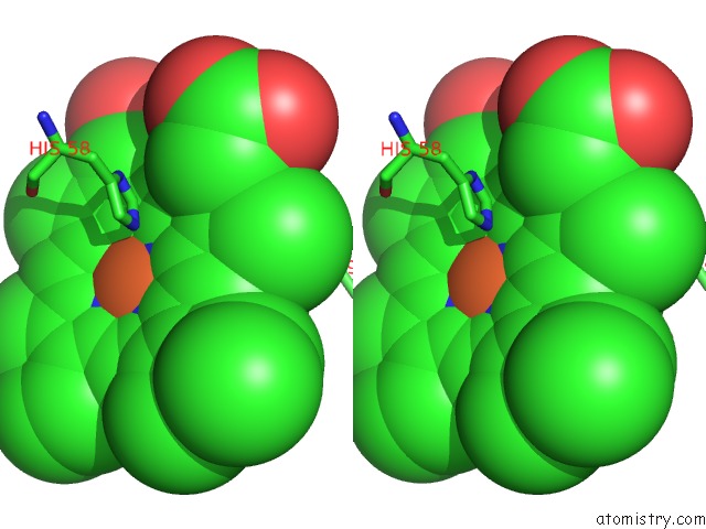
Iron binding site 1 out of 4 in 1gbv
Go back to
Iron binding site 1 out
of 4 in the (Alpha-Oxy, Beta-(C112G)Deoxy) T-State Human Hemoglobin
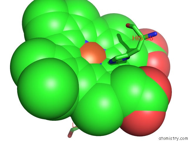
Mono view
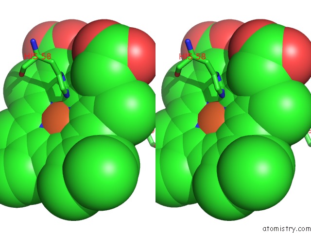
Stereo pair view
Mono view
Stereo pair view
A full contact list of Iron with other atoms in the Fe binding
site number 1 of (Alpha-Oxy, Beta-(C112G)Deoxy) T-State Human Hemoglobin within 5.0Å range:
|
Iron binding site 2 out of 4 in 1gbv
Go back to
Iron binding site 2 out
of 4 in the (Alpha-Oxy, Beta-(C112G)Deoxy) T-State Human Hemoglobin
Mono view

Stereo pair view
Mono view
Stereo pair view
A full contact list of Iron with other atoms in the Fe binding
site number 2 of (Alpha-Oxy, Beta-(C112G)Deoxy) T-State Human Hemoglobin within 5.0Å range:
|
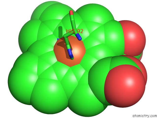
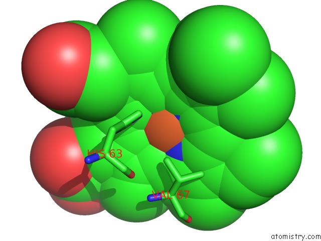
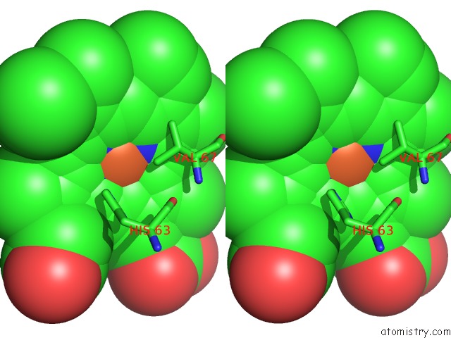
Iron binding site 3 out of 4 in 1gbv
Go back to
Iron binding site 3 out
of 4 in the (Alpha-Oxy, Beta-(C112G)Deoxy) T-State Human Hemoglobin
Mono view
Stereo pair view
Mono view
Stereo pair view
A full contact list of Iron with other atoms in the Fe binding
site number 3 of (Alpha-Oxy, Beta-(C112G)Deoxy) T-State Human Hemoglobin within 5.0Å range:
|
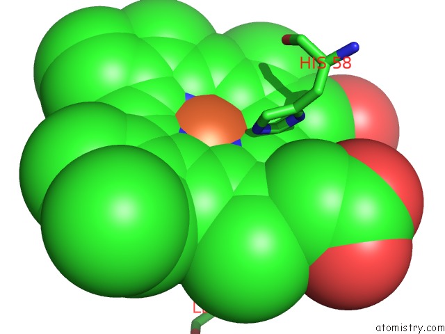
Iron binding site 4 out of 4 in 1gbv
Go back to
Iron binding site 4 out
of 4 in the (Alpha-Oxy, Beta-(C112G)Deoxy) T-State Human Hemoglobin
Mono view
Stereo pair view
Mono view
Stereo pair view
A full contact list of Iron with other atoms in the Fe binding
site number 4 of (Alpha-Oxy, Beta-(C112G)Deoxy) T-State Human Hemoglobin within 5.0Å range:
|
Reference:
G.B.Vasquez,
M.Karavitis,
X.Ji,
I.Pechik,
W.S.Brinigar,
G.L.Gilliland,
C.Fronticelli.
Cysteines BETA93 and BETA112 As Probes of Conformational and Functional Events at the Human Hemoglobin Subunit Interfaces. Biophys.J. V. 76 88 1999.
ISSN: ISSN 0006-3495
PubMed: 9876125
Page generated: Wed Jul 16 14:54:36 2025
ISSN: ISSN 0006-3495
PubMed: 9876125
Last articles
Fe in 2YXOFe in 2YRS
Fe in 2YXC
Fe in 2YNM
Fe in 2YVJ
Fe in 2YP1
Fe in 2YU2
Fe in 2YU1
Fe in 2YQB
Fe in 2YOO