Iron »
PDB 1gwf-1h5f »
1gy9 »
Iron in PDB 1gy9: Taurine/Alpha-Ketoglutarate Dioxygenase From Escherichia Coli
Enzymatic activity of Taurine/Alpha-Ketoglutarate Dioxygenase From Escherichia Coli
All present enzymatic activity of Taurine/Alpha-Ketoglutarate Dioxygenase From Escherichia Coli:
1.14.11.17;
1.14.11.17;
Protein crystallography data
The structure of Taurine/Alpha-Ketoglutarate Dioxygenase From Escherichia Coli, PDB code: 1gy9
was solved by
J.M.Elkins,
N.I.Burzlaff,
M.J.Ryle,
J.S.Lloyd,
I.J.Clifton,
J.E.Baldwin,
R.P.Hausinger,
P.L.Roach,
with X-Ray Crystallography technique. A brief refinement statistics is given in the table below:
| Resolution Low / High (Å) | 45.18 / 2.50 |
| Space group | P 62 2 2 |
| Cell size a, b, c (Å), α, β, γ (°) | 116.786, 116.786, 201.937, 90.00, 90.00, 120.00 |
| R / Rfree (%) | 26.1 / 29.3 |
Iron Binding Sites:
The binding sites of Iron atom in the Taurine/Alpha-Ketoglutarate Dioxygenase From Escherichia Coli
(pdb code 1gy9). This binding sites where shown within
5.0 Angstroms radius around Iron atom.
In total 3 binding sites of Iron where determined in the Taurine/Alpha-Ketoglutarate Dioxygenase From Escherichia Coli, PDB code: 1gy9:
Jump to Iron binding site number: 1; 2; 3;
In total 3 binding sites of Iron where determined in the Taurine/Alpha-Ketoglutarate Dioxygenase From Escherichia Coli, PDB code: 1gy9:
Jump to Iron binding site number: 1; 2; 3;
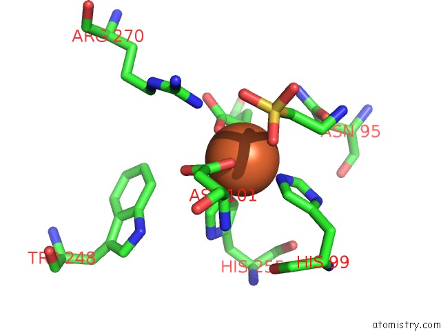
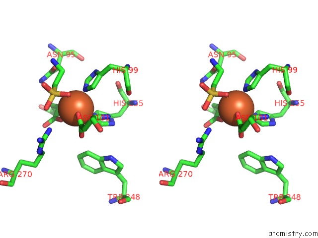
Iron binding site 1 out of 3 in 1gy9
Go back to
Iron binding site 1 out
of 3 in the Taurine/Alpha-Ketoglutarate Dioxygenase From Escherichia Coli
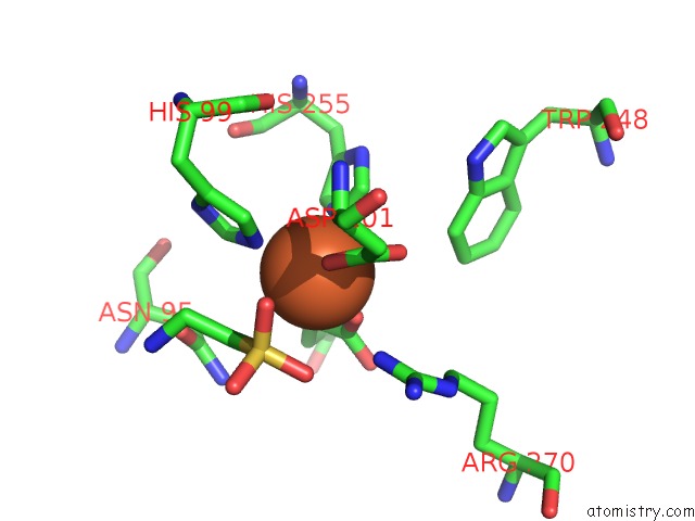
Mono view
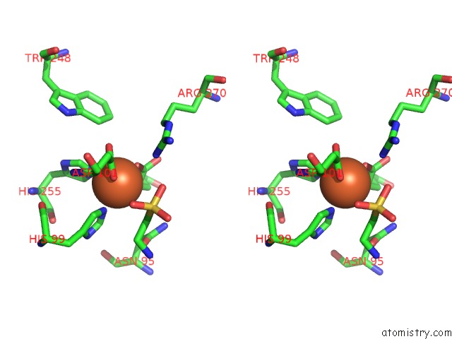
Stereo pair view
Mono view
Stereo pair view
A full contact list of Iron with other atoms in the Fe binding
site number 1 of Taurine/Alpha-Ketoglutarate Dioxygenase From Escherichia Coli within 5.0Å range:
|
Iron binding site 2 out of 3 in 1gy9
Go back to
Iron binding site 2 out
of 3 in the Taurine/Alpha-Ketoglutarate Dioxygenase From Escherichia Coli
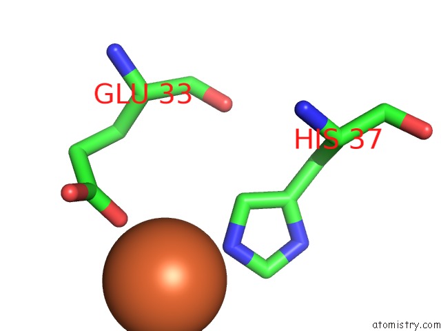
Mono view
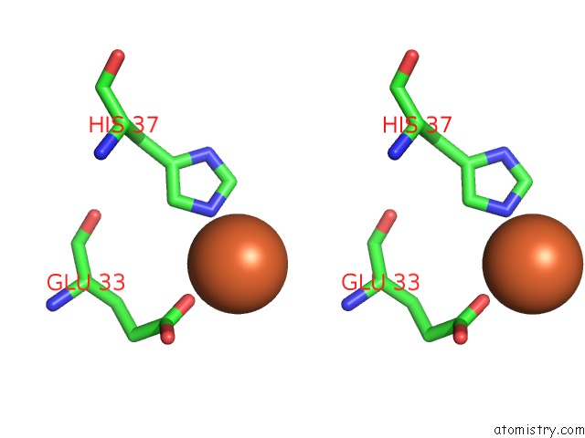
Stereo pair view
Mono view
Stereo pair view
A full contact list of Iron with other atoms in the Fe binding
site number 2 of Taurine/Alpha-Ketoglutarate Dioxygenase From Escherichia Coli within 5.0Å range:
|
Iron binding site 3 out of 3 in 1gy9
Go back to
Iron binding site 3 out
of 3 in the Taurine/Alpha-Ketoglutarate Dioxygenase From Escherichia Coli
Mono view
Stereo pair view
Mono view
Stereo pair view
A full contact list of Iron with other atoms in the Fe binding
site number 3 of Taurine/Alpha-Ketoglutarate Dioxygenase From Escherichia Coli within 5.0Å range:
|
Reference:
J.M.Elkins,
M.J.Ryle,
I.J.Clifton,
J.C.Dunning Hotopp,
J.S.Lloyd,
N.I.Burzlaff,
J.E.Baldwin,
R.P.Hausinger,
P.L.Roach.
X-Ray Crystal Structure of Escherichia Coli Taurine/Alpha-Ketoglutarate Dioxygenase Complexed to Ferrous Iron and Substrates. Biochemistry V. 41 5185 2002.
ISSN: ISSN 0006-2960
PubMed: 11955067
DOI: 10.1021/BI016014E
Page generated: Sat Aug 3 06:45:06 2024
ISSN: ISSN 0006-2960
PubMed: 11955067
DOI: 10.1021/BI016014E
Last articles
Zn in 9J0NZn in 9J0O
Zn in 9J0P
Zn in 9FJX
Zn in 9EKB
Zn in 9C0F
Zn in 9CAH
Zn in 9CH0
Zn in 9CH3
Zn in 9CH1