Iron in PDB 1h2r: Three-Dimensional Structure of Ni-Fe Hydrogenase From Desulfivibrio Vulgaris Miyazaki F in the Reduced Form at 1.4 A Resolution
Enzymatic activity of Three-Dimensional Structure of Ni-Fe Hydrogenase From Desulfivibrio Vulgaris Miyazaki F in the Reduced Form at 1.4 A Resolution
All present enzymatic activity of Three-Dimensional Structure of Ni-Fe Hydrogenase From Desulfivibrio Vulgaris Miyazaki F in the Reduced Form at 1.4 A Resolution:
1.12.2.1;
Protein crystallography data
The structure of Three-Dimensional Structure of Ni-Fe Hydrogenase From Desulfivibrio Vulgaris Miyazaki F in the Reduced Form at 1.4 A Resolution, PDB code: 1h2r
was solved by
Y.Higuchi,
H.Ogata,
with X-Ray Crystallography technique. A brief refinement statistics is given in the table below:
Resolution Low / High (Å)
|
6.00 /
1.40
|
Space group
|
P 21 21 21
|
Cell size a, b, c (Å), α, β, γ (°)
|
100.440,
126.860,
66.680,
90.00,
90.00,
90.00
|
R / Rfree (%)
|
21.8 /
25.4
|
Other elements in 1h2r:
The structure of Three-Dimensional Structure of Ni-Fe Hydrogenase From Desulfivibrio Vulgaris Miyazaki F in the Reduced Form at 1.4 A Resolution also contains other interesting chemical elements:
Iron Binding Sites:
Pages:
>>> Page 1 <<<
Page 2, Binding sites: 11 -
12;
Binding sites:
The binding sites of Iron atom in the Three-Dimensional Structure of Ni-Fe Hydrogenase From Desulfivibrio Vulgaris Miyazaki F in the Reduced Form at 1.4 A Resolution
(pdb code 1h2r). This binding sites where shown within
5.0 Angstroms radius around Iron atom.
In total 12 binding sites of Iron where determined in the
Three-Dimensional Structure of Ni-Fe Hydrogenase From Desulfivibrio Vulgaris Miyazaki F in the Reduced Form at 1.4 A Resolution, PDB code: 1h2r:
Jump to Iron binding site number:
1;
2;
3;
4;
5;
6;
7;
8;
9;
10;
Iron binding site 1 out
of 12 in 1h2r
Go back to
Iron Binding Sites List in 1h2r
Iron binding site 1 out
of 12 in the Three-Dimensional Structure of Ni-Fe Hydrogenase From Desulfivibrio Vulgaris Miyazaki F in the Reduced Form at 1.4 A Resolution
 Mono view
 Stereo pair view
|
A full contact list of Iron with other atoms in the Fe binding
site number 1 of Three-Dimensional Structure of Ni-Fe Hydrogenase From Desulfivibrio Vulgaris Miyazaki F in the Reduced Form at 1.4 A Resolution within 5.0Å range:
probe
|
atom
|
residue
|
distance (Å)
|
B
|
Occ
|
S:Fe1001
b:10.2
occ:1.00
|
FE1
|
S:SF41001
|
0.0
|
10.2
|
1.0
|
S4
|
S:SF41001
|
2.3
|
10.9
|
1.0
|
SG
|
S:CYS17
|
2.3
|
10.4
|
1.0
|
S3
|
S:SF41001
|
2.3
|
12.6
|
1.0
|
S2
|
S:SF41001
|
2.3
|
11.3
|
1.0
|
FE3
|
S:SF41001
|
2.7
|
11.3
|
1.0
|
FE2
|
S:SF41001
|
2.7
|
11.0
|
1.0
|
FE4
|
S:SF41001
|
2.7
|
11.6
|
1.0
|
CB
|
S:CYS17
|
3.4
|
11.9
|
1.0
|
N
|
S:CYS17
|
3.8
|
10.7
|
1.0
|
S1
|
S:SF41001
|
3.9
|
11.2
|
1.0
|
CA
|
S:CYS17
|
4.1
|
10.6
|
1.0
|
CE1
|
L:HIS235
|
4.2
|
8.3
|
1.0
|
N
|
S:GLY19
|
4.3
|
9.8
|
1.0
|
O
|
L:HOH3009
|
4.5
|
9.5
|
1.0
|
CG
|
L:ARG79
|
4.6
|
15.9
|
1.0
|
CA
|
S:GLY19
|
4.6
|
9.5
|
1.0
|
N
|
S:THR18
|
4.6
|
10.8
|
1.0
|
C
|
S:CYS17
|
4.6
|
10.9
|
1.0
|
N
|
S:CYS20
|
4.7
|
10.2
|
1.0
|
SG
|
S:CYS150
|
4.8
|
10.4
|
1.0
|
SG
|
S:CYS114
|
4.8
|
10.3
|
1.0
|
SG
|
S:CYS20
|
4.8
|
12.3
|
1.0
|
CD
|
L:ARG79
|
4.9
|
19.3
|
1.0
|
C
|
S:GLU16
|
4.9
|
11.0
|
1.0
|
ND1
|
L:HIS235
|
4.9
|
11.8
|
1.0
|
O
|
L:ARG79
|
5.0
|
10.3
|
1.0
|
|
Iron binding site 2 out
of 12 in 1h2r
Go back to
Iron Binding Sites List in 1h2r
Iron binding site 2 out
of 12 in the Three-Dimensional Structure of Ni-Fe Hydrogenase From Desulfivibrio Vulgaris Miyazaki F in the Reduced Form at 1.4 A Resolution
 Mono view
 Stereo pair view
|
A full contact list of Iron with other atoms in the Fe binding
site number 2 of Three-Dimensional Structure of Ni-Fe Hydrogenase From Desulfivibrio Vulgaris Miyazaki F in the Reduced Form at 1.4 A Resolution within 5.0Å range:
probe
|
atom
|
residue
|
distance (Å)
|
B
|
Occ
|
S:Fe1001
b:11.0
occ:1.00
|
FE2
|
S:SF41001
|
0.0
|
11.0
|
1.0
|
S4
|
S:SF41001
|
2.3
|
10.9
|
1.0
|
SG
|
S:CYS114
|
2.3
|
10.3
|
1.0
|
S3
|
S:SF41001
|
2.3
|
12.6
|
1.0
|
S1
|
S:SF41001
|
2.3
|
11.2
|
1.0
|
FE3
|
S:SF41001
|
2.6
|
11.3
|
1.0
|
FE1
|
S:SF41001
|
2.7
|
10.2
|
1.0
|
FE4
|
S:SF41001
|
2.8
|
11.6
|
1.0
|
CB
|
S:CYS114
|
3.2
|
9.8
|
1.0
|
S2
|
S:SF41001
|
3.9
|
11.3
|
1.0
|
O
|
L:HOH3009
|
4.1
|
9.5
|
1.0
|
O
|
S:HOH3425
|
4.1
|
10.4
|
1.0
|
N
|
S:CYS114
|
4.1
|
10.4
|
1.0
|
O
|
S:HOH3080
|
4.1
|
10.5
|
1.0
|
CA
|
S:CYS114
|
4.2
|
11.0
|
1.0
|
O
|
S:HOH3034
|
4.3
|
87.8
|
1.0
|
SG
|
S:CYS150
|
4.5
|
10.4
|
1.0
|
SG
|
S:CYS20
|
4.8
|
12.3
|
1.0
|
SG
|
S:CYS17
|
4.9
|
10.4
|
1.0
|
N
|
S:CYS17
|
4.9
|
10.7
|
1.0
|
CA
|
S:GLU16
|
5.0
|
11.1
|
1.0
|
|
Iron binding site 3 out
of 12 in 1h2r
Go back to
Iron Binding Sites List in 1h2r
Iron binding site 3 out
of 12 in the Three-Dimensional Structure of Ni-Fe Hydrogenase From Desulfivibrio Vulgaris Miyazaki F in the Reduced Form at 1.4 A Resolution
 Mono view
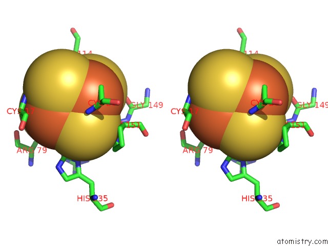
 Stereo pair view
|
A full contact list of Iron with other atoms in the Fe binding
site number 3 of Three-Dimensional Structure of Ni-Fe Hydrogenase From Desulfivibrio Vulgaris Miyazaki F in the Reduced Form at 1.4 A Resolution within 5.0Å range:
probe
|
atom
|
residue
|
distance (Å)
|
B
|
Occ
|
S:Fe1001
b:11.3
occ:1.00
|
FE3
|
S:SF41001
|
0.0
|
11.3
|
1.0
|
S4
|
S:SF41001
|
2.3
|
10.9
|
1.0
|
SG
|
S:CYS150
|
2.3
|
10.4
|
1.0
|
S2
|
S:SF41001
|
2.3
|
11.3
|
1.0
|
S1
|
S:SF41001
|
2.3
|
11.2
|
1.0
|
FE2
|
S:SF41001
|
2.6
|
11.0
|
1.0
|
FE1
|
S:SF41001
|
2.7
|
10.2
|
1.0
|
FE4
|
S:SF41001
|
2.7
|
11.6
|
1.0
|
CB
|
S:CYS150
|
3.5
|
9.6
|
1.0
|
CA
|
S:CYS150
|
3.6
|
10.6
|
1.0
|
S3
|
S:SF41001
|
3.9
|
12.6
|
1.0
|
C
|
S:CYS150
|
3.9
|
10.1
|
1.0
|
CD
|
L:ARG79
|
4.0
|
19.3
|
1.0
|
CG
|
L:ARG79
|
4.0
|
15.9
|
1.0
|
O
|
S:HOH3425
|
4.0
|
10.4
|
1.0
|
O
|
S:CYS150
|
4.3
|
9.9
|
1.0
|
N
|
S:PRO151
|
4.4
|
9.9
|
1.0
|
SG
|
S:CYS114
|
4.4
|
10.3
|
1.0
|
CE1
|
L:HIS235
|
4.6
|
8.3
|
1.0
|
SG
|
S:CYS17
|
4.6
|
10.4
|
1.0
|
ND1
|
L:HIS235
|
4.7
|
11.8
|
1.0
|
O
|
S:GLY149
|
4.7
|
8.6
|
1.0
|
CA
|
S:PRO151
|
4.7
|
9.4
|
1.0
|
SG
|
S:CYS20
|
4.8
|
12.3
|
1.0
|
N
|
S:CYS150
|
4.9
|
9.5
|
1.0
|
|
Iron binding site 4 out
of 12 in 1h2r
Go back to
Iron Binding Sites List in 1h2r
Iron binding site 4 out
of 12 in the Three-Dimensional Structure of Ni-Fe Hydrogenase From Desulfivibrio Vulgaris Miyazaki F in the Reduced Form at 1.4 A Resolution
 Mono view
 Stereo pair view
|
A full contact list of Iron with other atoms in the Fe binding
site number 4 of Three-Dimensional Structure of Ni-Fe Hydrogenase From Desulfivibrio Vulgaris Miyazaki F in the Reduced Form at 1.4 A Resolution within 5.0Å range:
probe
|
atom
|
residue
|
distance (Å)
|
B
|
Occ
|
S:Fe1001
b:11.6
occ:1.00
|
FE4
|
S:SF41001
|
0.0
|
11.6
|
1.0
|
SG
|
S:CYS20
|
2.3
|
12.3
|
1.0
|
S2
|
S:SF41001
|
2.3
|
11.3
|
1.0
|
S1
|
S:SF41001
|
2.3
|
11.2
|
1.0
|
S3
|
S:SF41001
|
2.4
|
12.6
|
1.0
|
FE1
|
S:SF41001
|
2.7
|
10.2
|
1.0
|
FE3
|
S:SF41001
|
2.7
|
11.3
|
1.0
|
FE2
|
S:SF41001
|
2.8
|
11.0
|
1.0
|
CB
|
S:CYS20
|
3.5
|
10.4
|
1.0
|
N
|
S:CYS20
|
3.8
|
10.2
|
1.0
|
O
|
S:HOH3080
|
4.0
|
10.5
|
1.0
|
S4
|
S:SF41001
|
4.0
|
10.9
|
1.0
|
CA
|
S:CYS20
|
4.2
|
11.2
|
1.0
|
CA
|
S:PRO151
|
4.4
|
9.4
|
1.0
|
CB
|
S:PRO151
|
4.4
|
9.0
|
1.0
|
CA
|
S:GLY112
|
4.6
|
10.1
|
1.0
|
C
|
S:GLY19
|
4.6
|
9.3
|
1.0
|
OE2
|
S:GLU75
|
4.7
|
14.6
|
1.0
|
SG
|
S:CYS17
|
4.7
|
10.4
|
1.0
|
O
|
S:GLY149
|
4.8
|
8.6
|
1.0
|
CA
|
S:CYS150
|
4.8
|
10.6
|
1.0
|
N
|
S:PRO151
|
4.8
|
9.9
|
1.0
|
SG
|
S:CYS150
|
4.9
|
10.4
|
1.0
|
SG
|
S:CYS114
|
5.0
|
10.3
|
1.0
|
CA
|
S:GLY19
|
5.0
|
9.5
|
1.0
|
N
|
S:GLY19
|
5.0
|
9.8
|
1.0
|
|
Iron binding site 5 out
of 12 in 1h2r
Go back to
Iron Binding Sites List in 1h2r
Iron binding site 5 out
of 12 in the Three-Dimensional Structure of Ni-Fe Hydrogenase From Desulfivibrio Vulgaris Miyazaki F in the Reduced Form at 1.4 A Resolution
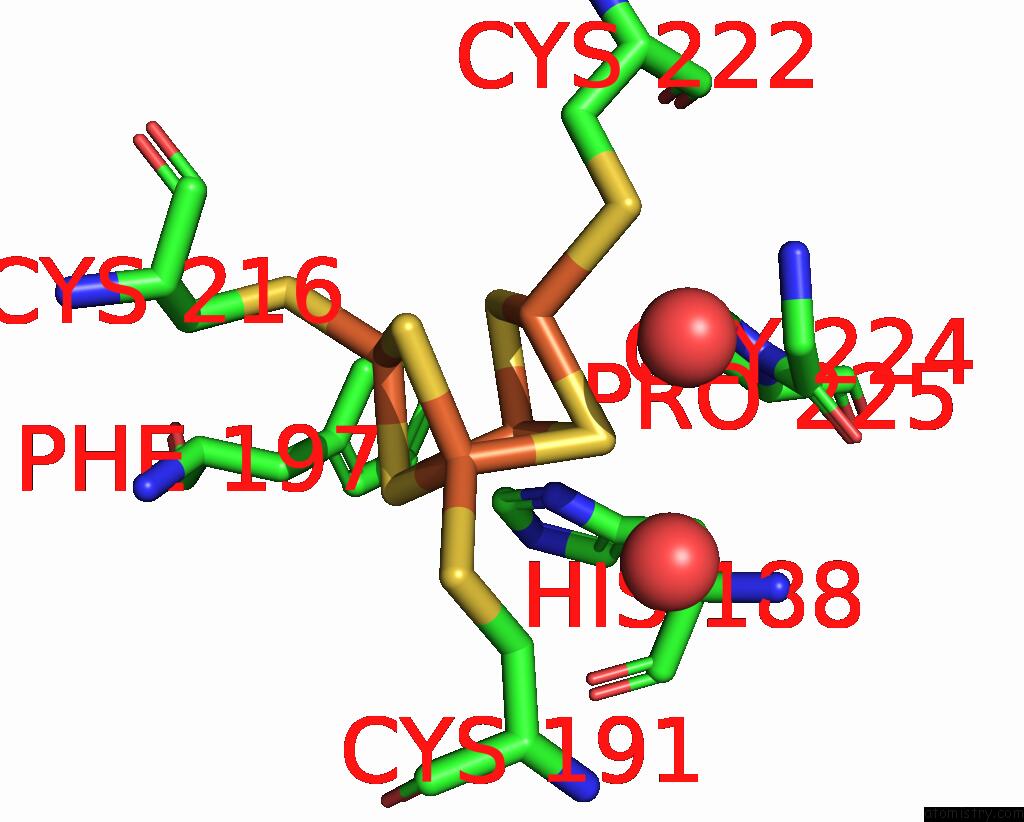
 Mono view
 Stereo pair view
|
A full contact list of Iron with other atoms in the Fe binding
site number 5 of Three-Dimensional Structure of Ni-Fe Hydrogenase From Desulfivibrio Vulgaris Miyazaki F in the Reduced Form at 1.4 A Resolution within 5.0Å range:
probe
|
atom
|
residue
|
distance (Å)
|
B
|
Occ
|
S:Fe1002
b:12.8
occ:1.00
|
FE1
|
S:SF41002
|
0.0
|
12.8
|
1.0
|
ND1
|
S:HIS188
|
2.1
|
12.7
|
1.0
|
S4
|
S:SF41002
|
2.3
|
13.3
|
1.0
|
S3
|
S:SF41002
|
2.3
|
13.0
|
1.0
|
S2
|
S:SF41002
|
2.3
|
14.0
|
1.0
|
FE2
|
S:SF41002
|
2.7
|
12.1
|
1.0
|
FE4
|
S:SF41002
|
2.7
|
13.3
|
1.0
|
FE3
|
S:SF41002
|
2.7
|
11.9
|
1.0
|
CE1
|
S:HIS188
|
2.9
|
14.6
|
1.0
|
CG
|
S:HIS188
|
3.2
|
13.2
|
1.0
|
CB
|
S:HIS188
|
3.7
|
12.0
|
1.0
|
S1
|
S:SF41002
|
3.9
|
13.3
|
1.0
|
CA
|
S:HIS188
|
3.9
|
13.4
|
1.0
|
NE2
|
S:HIS188
|
4.1
|
13.4
|
1.0
|
CD2
|
S:HIS188
|
4.3
|
12.6
|
1.0
|
CD
|
S:PRO225
|
4.3
|
12.4
|
1.0
|
CG
|
S:PRO225
|
4.3
|
13.1
|
1.0
|
O
|
S:HIS188
|
4.5
|
13.9
|
1.0
|
CB
|
S:CYS191
|
4.5
|
14.1
|
1.0
|
O
|
S:HOH3338
|
4.7
|
80.0
|
1.0
|
C
|
S:HIS188
|
4.7
|
13.1
|
1.0
|
SG
|
S:CYS191
|
4.7
|
14.2
|
1.0
|
SG
|
S:CYS216
|
4.7
|
13.9
|
1.0
|
CD2
|
S:PHE197
|
4.8
|
17.8
|
1.0
|
SG
|
S:CYS222
|
4.8
|
11.6
|
1.0
|
O
|
S:HOH3012
|
4.9
|
96.2
|
1.0
|
N
|
S:PRO225
|
4.9
|
12.2
|
1.0
|
CA
|
S:GLY224
|
5.0
|
11.1
|
1.0
|
|
Iron binding site 6 out
of 12 in 1h2r
Go back to
Iron Binding Sites List in 1h2r
Iron binding site 6 out
of 12 in the Three-Dimensional Structure of Ni-Fe Hydrogenase From Desulfivibrio Vulgaris Miyazaki F in the Reduced Form at 1.4 A Resolution
 Mono view
 Stereo pair view
|
A full contact list of Iron with other atoms in the Fe binding
site number 6 of Three-Dimensional Structure of Ni-Fe Hydrogenase From Desulfivibrio Vulgaris Miyazaki F in the Reduced Form at 1.4 A Resolution within 5.0Å range:
probe
|
atom
|
residue
|
distance (Å)
|
B
|
Occ
|
S:Fe1002
b:12.1
occ:1.00
|
FE2
|
S:SF41002
|
0.0
|
12.1
|
1.0
|
S3
|
S:SF41002
|
2.3
|
13.0
|
1.0
|
S4
|
S:SF41002
|
2.3
|
13.3
|
1.0
|
SG
|
S:CYS222
|
2.3
|
11.6
|
1.0
|
S1
|
S:SF41002
|
2.4
|
13.3
|
1.0
|
FE1
|
S:SF41002
|
2.7
|
12.8
|
1.0
|
FE3
|
S:SF41002
|
2.7
|
11.9
|
1.0
|
FE4
|
S:SF41002
|
2.7
|
13.3
|
1.0
|
O
|
S:HOH3012
|
3.1
|
96.2
|
1.0
|
CB
|
S:CYS222
|
3.3
|
11.4
|
1.0
|
S2
|
S:SF41002
|
3.9
|
14.0
|
1.0
|
CA
|
S:GLY224
|
4.6
|
11.1
|
1.0
|
N
|
S:GLY224
|
4.6
|
12.4
|
1.0
|
ND1
|
S:HIS188
|
4.6
|
12.7
|
1.0
|
CD
|
S:PRO225
|
4.7
|
12.4
|
1.0
|
CG1
|
S:VAL243
|
4.7
|
19.0
|
1.0
|
CA
|
S:CYS222
|
4.7
|
11.0
|
1.0
|
SG
|
S:CYS216
|
4.7
|
13.9
|
1.0
|
O
|
S:HOH3338
|
4.8
|
80.0
|
1.0
|
SG
|
S:CYS191
|
4.8
|
14.2
|
1.0
|
N
|
S:LEU217
|
4.8
|
13.8
|
1.0
|
N
|
S:TYR218
|
4.9
|
13.3
|
1.0
|
C
|
S:CYS222
|
5.0
|
10.5
|
1.0
|
CB
|
S:LEU217
|
5.0
|
13.2
|
1.0
|
C
|
S:LEU217
|
5.0
|
13.9
|
1.0
|
|
Iron binding site 7 out
of 12 in 1h2r
Go back to
Iron Binding Sites List in 1h2r
Iron binding site 7 out
of 12 in the Three-Dimensional Structure of Ni-Fe Hydrogenase From Desulfivibrio Vulgaris Miyazaki F in the Reduced Form at 1.4 A Resolution
 Mono view
 Stereo pair view
|
A full contact list of Iron with other atoms in the Fe binding
site number 7 of Three-Dimensional Structure of Ni-Fe Hydrogenase From Desulfivibrio Vulgaris Miyazaki F in the Reduced Form at 1.4 A Resolution within 5.0Å range:
probe
|
atom
|
residue
|
distance (Å)
|
B
|
Occ
|
S:Fe1002
b:11.9
occ:1.00
|
FE3
|
S:SF41002
|
0.0
|
11.9
|
1.0
|
S1
|
S:SF41002
|
2.3
|
13.3
|
1.0
|
SG
|
S:CYS191
|
2.3
|
14.2
|
1.0
|
S2
|
S:SF41002
|
2.3
|
14.0
|
1.0
|
S4
|
S:SF41002
|
2.3
|
13.3
|
1.0
|
FE2
|
S:SF41002
|
2.7
|
12.1
|
1.0
|
FE1
|
S:SF41002
|
2.7
|
12.8
|
1.0
|
FE4
|
S:SF41002
|
2.8
|
13.3
|
1.0
|
CB
|
S:CYS191
|
3.1
|
14.1
|
1.0
|
O
|
S:HOH3338
|
3.7
|
80.0
|
1.0
|
S3
|
S:SF41002
|
4.0
|
13.0
|
1.0
|
CB
|
S:ARG193
|
4.1
|
15.7
|
1.0
|
O
|
S:HOH3012
|
4.2
|
96.2
|
1.0
|
ND1
|
S:HIS188
|
4.5
|
12.7
|
1.0
|
CG2
|
S:VAL243
|
4.5
|
21.1
|
1.0
|
CA
|
S:CYS191
|
4.6
|
14.7
|
1.0
|
C
|
S:ARG193
|
4.7
|
17.1
|
1.0
|
CA
|
S:ARG193
|
4.7
|
16.3
|
1.0
|
N
|
S:ARG193
|
4.7
|
15.7
|
1.0
|
N
|
S:LEU194
|
4.7
|
18.1
|
1.0
|
CG1
|
S:VAL243
|
4.8
|
19.0
|
1.0
|
CG
|
S:ARG193
|
4.8
|
14.1
|
1.0
|
SG
|
S:CYS222
|
4.8
|
11.6
|
1.0
|
CH2
|
S:TRP241
|
4.9
|
20.9
|
1.0
|
CD
|
S:ARG193
|
5.0
|
14.3
|
1.0
|
SG
|
S:CYS216
|
5.0
|
13.9
|
1.0
|
|
Iron binding site 8 out
of 12 in 1h2r
Go back to
Iron Binding Sites List in 1h2r
Iron binding site 8 out
of 12 in the Three-Dimensional Structure of Ni-Fe Hydrogenase From Desulfivibrio Vulgaris Miyazaki F in the Reduced Form at 1.4 A Resolution
 Mono view
 Stereo pair view
|
A full contact list of Iron with other atoms in the Fe binding
site number 8 of Three-Dimensional Structure of Ni-Fe Hydrogenase From Desulfivibrio Vulgaris Miyazaki F in the Reduced Form at 1.4 A Resolution within 5.0Å range:
probe
|
atom
|
residue
|
distance (Å)
|
B
|
Occ
|
S:Fe1002
b:13.3
occ:1.00
|
FE4
|
S:SF41002
|
0.0
|
13.3
|
1.0
|
S2
|
S:SF41002
|
2.3
|
14.0
|
1.0
|
SG
|
S:CYS216
|
2.3
|
13.9
|
1.0
|
S1
|
S:SF41002
|
2.3
|
13.3
|
1.0
|
S3
|
S:SF41002
|
2.4
|
13.0
|
1.0
|
FE1
|
S:SF41002
|
2.7
|
12.8
|
1.0
|
FE2
|
S:SF41002
|
2.7
|
12.1
|
1.0
|
FE3
|
S:SF41002
|
2.8
|
11.9
|
1.0
|
CB
|
S:CYS216
|
3.4
|
13.1
|
1.0
|
N
|
S:LEU217
|
3.8
|
13.8
|
1.0
|
CA
|
S:CYS216
|
3.9
|
14.2
|
1.0
|
S4
|
S:SF41002
|
3.9
|
13.3
|
1.0
|
N
|
S:TYR218
|
4.2
|
13.3
|
1.0
|
C
|
S:CYS216
|
4.3
|
13.8
|
1.0
|
CB
|
S:PHE197
|
4.4
|
20.3
|
1.0
|
ND1
|
S:HIS188
|
4.5
|
12.7
|
1.0
|
CD2
|
S:PHE197
|
4.5
|
17.8
|
1.0
|
CE1
|
S:HIS188
|
4.7
|
14.6
|
1.0
|
CB
|
S:ARG193
|
4.7
|
15.7
|
1.0
|
CA
|
S:LEU217
|
4.7
|
13.7
|
1.0
|
CB
|
S:TYR218
|
4.7
|
14.9
|
1.0
|
C
|
S:LEU217
|
4.8
|
13.9
|
1.0
|
CG
|
S:PHE197
|
4.8
|
17.9
|
1.0
|
SG
|
S:CYS222
|
4.9
|
11.6
|
1.0
|
O
|
S:ARG193
|
4.9
|
16.3
|
1.0
|
SG
|
S:CYS191
|
4.9
|
14.2
|
1.0
|
CA
|
S:TYR218
|
4.9
|
14.7
|
1.0
|
|
Iron binding site 9 out
of 12 in 1h2r
Go back to
Iron Binding Sites List in 1h2r
Iron binding site 9 out
of 12 in the Three-Dimensional Structure of Ni-Fe Hydrogenase From Desulfivibrio Vulgaris Miyazaki F in the Reduced Form at 1.4 A Resolution
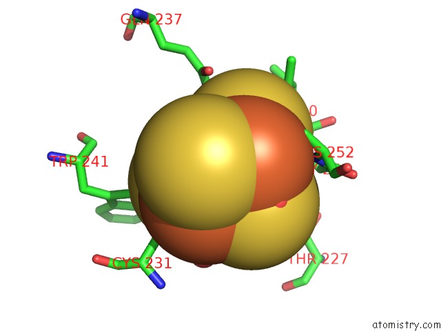
 Mono view
 Stereo pair view
|
A full contact list of Iron with other atoms in the Fe binding
site number 9 of Three-Dimensional Structure of Ni-Fe Hydrogenase From Desulfivibrio Vulgaris Miyazaki F in the Reduced Form at 1.4 A Resolution within 5.0Å range:
probe
|
atom
|
residue
|
distance (Å)
|
B
|
Occ
|
S:Fe1003
b:11.2
occ:1.00
|
FE1
|
S:F3S1003
|
0.0
|
11.2
|
1.0
|
S3
|
S:F3S1003
|
2.3
|
12.6
|
1.0
|
S1
|
S:F3S1003
|
2.3
|
12.8
|
1.0
|
S2
|
S:F3S1003
|
2.3
|
12.3
|
1.0
|
SG
|
S:CYS249
|
2.4
|
11.5
|
1.0
|
FE4
|
S:F3S1003
|
2.7
|
11.1
|
1.0
|
FE3
|
S:F3S1003
|
2.7
|
11.8
|
1.0
|
CB
|
S:CYS249
|
3.4
|
11.5
|
1.0
|
O
|
S:HOH3184
|
3.4
|
0.0
|
1.0
|
CA
|
S:CYS249
|
3.8
|
10.9
|
1.0
|
S4
|
S:F3S1003
|
4.0
|
10.8
|
1.0
|
N
|
S:ILE250
|
4.1
|
10.8
|
1.0
|
N
|
S:GLY251
|
4.2
|
10.4
|
1.0
|
C
|
S:CYS249
|
4.3
|
10.7
|
1.0
|
N
|
S:CYS252
|
4.6
|
11.7
|
1.0
|
O
|
S:HOH3067
|
4.7
|
58.8
|
1.0
|
CA
|
S:GLY251
|
4.8
|
10.6
|
1.0
|
SG
|
S:CYS252
|
4.8
|
12.0
|
1.0
|
CG2
|
S:THR227
|
4.8
|
10.9
|
1.0
|
SG
|
S:CYS231
|
4.9
|
13.1
|
1.0
|
O
|
S:HOH3237
|
4.9
|
98.9
|
1.0
|
NE2
|
L:GLN237
|
4.9
|
12.0
|
1.0
|
NE1
|
S:TRP241
|
5.0
|
26.0
|
1.0
|
|
Iron binding site 10 out
of 12 in 1h2r
Go back to
Iron Binding Sites List in 1h2r
Iron binding site 10 out
of 12 in the Three-Dimensional Structure of Ni-Fe Hydrogenase From Desulfivibrio Vulgaris Miyazaki F in the Reduced Form at 1.4 A Resolution
 Mono view
 Stereo pair view
|
A full contact list of Iron with other atoms in the Fe binding
site number 10 of Three-Dimensional Structure of Ni-Fe Hydrogenase From Desulfivibrio Vulgaris Miyazaki F in the Reduced Form at 1.4 A Resolution within 5.0Å range:
probe
|
atom
|
residue
|
distance (Å)
|
B
|
Occ
|
S:Fe1003
b:11.8
occ:1.00
|
FE3
|
S:F3S1003
|
0.0
|
11.8
|
1.0
|
S1
|
S:F3S1003
|
2.3
|
12.8
|
1.0
|
S4
|
S:F3S1003
|
2.3
|
10.8
|
1.0
|
SG
|
S:CYS231
|
2.3
|
13.1
|
1.0
|
S3
|
S:F3S1003
|
2.4
|
12.6
|
1.0
|
FE4
|
S:F3S1003
|
2.7
|
11.1
|
1.0
|
FE1
|
S:F3S1003
|
2.7
|
11.2
|
1.0
|
CB
|
S:CYS231
|
3.4
|
12.9
|
1.0
|
O
|
S:HOH3067
|
3.5
|
58.8
|
1.0
|
O
|
S:HOH3184
|
3.8
|
0.0
|
1.0
|
OD1
|
S:ASN229
|
4.0
|
13.5
|
1.0
|
S2
|
S:F3S1003
|
4.0
|
12.3
|
1.0
|
NE1
|
S:TRP241
|
4.0
|
26.0
|
1.0
|
O
|
S:HOH3237
|
4.2
|
98.9
|
1.0
|
O
|
S:HOH3043
|
4.4
|
9.7
|
1.0
|
CD1
|
S:TRP241
|
4.4
|
25.6
|
1.0
|
CG
|
S:ASN229
|
4.5
|
12.3
|
1.0
|
CE2
|
S:PHE236
|
4.6
|
12.0
|
1.0
|
CB
|
S:ASN229
|
4.6
|
10.7
|
1.0
|
CZ
|
S:PHE236
|
4.7
|
13.5
|
1.0
|
SG
|
S:CYS252
|
4.8
|
12.0
|
1.0
|
CA
|
S:CYS231
|
4.8
|
13.7
|
1.0
|
CG
|
S:PRO242
|
4.8
|
13.9
|
1.0
|
SG
|
S:CYS249
|
4.9
|
11.5
|
1.0
|
CD
|
S:PRO242
|
5.0
|
14.2
|
1.0
|
|
Reference:
Y.Higuchi,
H.Ogata,
K.Miki,
N.Yasuoka,
T.Yagi.
Removal of the Bridging Ligand Atom at the Ni-Fe Active Site of [Nife] Hydrogenase Upon Reduction with H2, As Revealed By X-Ray Structure Analysis at 1.4 A Resolution. Structure Fold.Des. V. 7 549 1999.
ISSN: ISSN 0969-2126
PubMed: 10378274
DOI: 10.1016/S0969-2126(99)80071-9
Page generated: Sat Aug 3 06:52:26 2024
|