Iron »
PDB 1h5g-1hdb »
1h5k »
Iron in PDB 1h5k: X-Ray Induced Reduction of Horseradish Peroxidase C1A Compound III (78-89% Dose)
Enzymatic activity of X-Ray Induced Reduction of Horseradish Peroxidase C1A Compound III (78-89% Dose)
All present enzymatic activity of X-Ray Induced Reduction of Horseradish Peroxidase C1A Compound III (78-89% Dose):
1.11.1.7;
1.11.1.7;
Protein crystallography data
The structure of X-Ray Induced Reduction of Horseradish Peroxidase C1A Compound III (78-89% Dose), PDB code: 1h5k
was solved by
G.I.Berglund,
G.H.Carlsson,
J.Hajdu,
A.T.Smith,
H.Szoke,
A.Henriksen,
with X-Ray Crystallography technique. A brief refinement statistics is given in the table below:
| Resolution Low / High (Å) | 34.60 / 1.60 |
| Space group | P 21 21 21 |
| Cell size a, b, c (Å), α, β, γ (°) | 40.318, 67.392, 117.467, 90.00, 90.00, 90.00 |
| R / Rfree (%) | 19 / 21.3 |
Other elements in 1h5k:
The structure of X-Ray Induced Reduction of Horseradish Peroxidase C1A Compound III (78-89% Dose) also contains other interesting chemical elements:
| Calcium | (Ca) | 2 atoms |
Iron Binding Sites:
The binding sites of Iron atom in the X-Ray Induced Reduction of Horseradish Peroxidase C1A Compound III (78-89% Dose)
(pdb code 1h5k). This binding sites where shown within
5.0 Angstroms radius around Iron atom.
In total only one binding site of Iron was determined in the X-Ray Induced Reduction of Horseradish Peroxidase C1A Compound III (78-89% Dose), PDB code: 1h5k:
In total only one binding site of Iron was determined in the X-Ray Induced Reduction of Horseradish Peroxidase C1A Compound III (78-89% Dose), PDB code: 1h5k:
Iron binding site 1 out of 1 in 1h5k
Go back to
Iron binding site 1 out
of 1 in the X-Ray Induced Reduction of Horseradish Peroxidase C1A Compound III (78-89% Dose)
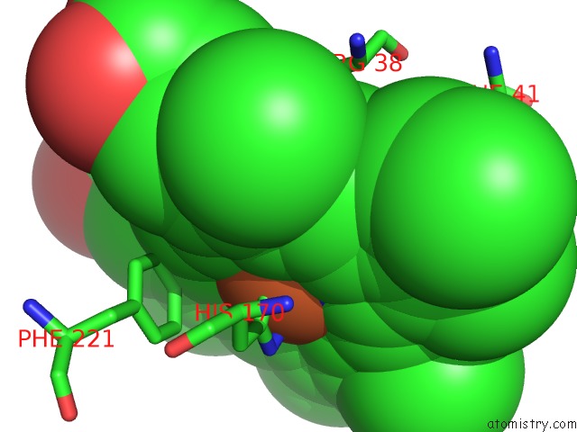
Mono view
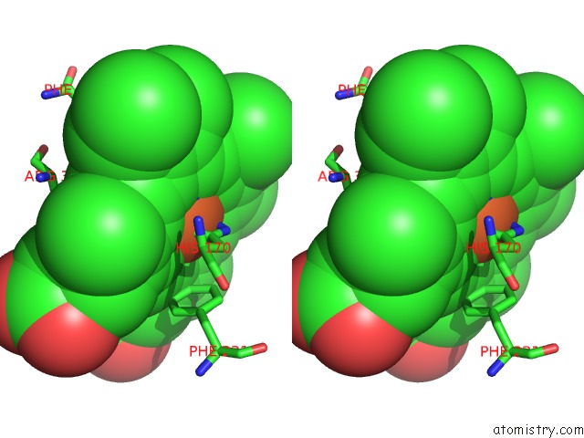
Stereo pair view
Mono view
Stereo pair view
A full contact list of Iron with other atoms in the Fe binding
site number 1 of X-Ray Induced Reduction of Horseradish Peroxidase C1A Compound III (78-89% Dose) within 5.0Å range:
|
Reference:
G.I.Berglund,
G.H.Carlsson,
A.T.Smith,
H.Szoke,
A.Henriksen,
J.Hajdu.
The Catalytic Pathway of Horseradish Peroxidase at High Resolution Nature V. 417 463 2002.
ISSN: ISSN 0028-0836
PubMed: 12024218
DOI: 10.1038/417463A
Page generated: Sat Aug 3 07:16:57 2024
ISSN: ISSN 0028-0836
PubMed: 12024218
DOI: 10.1038/417463A
Last articles
F in 4LB3F in 4LA6
F in 4L9I
F in 4L3O
F in 4L7H
F in 4L8M
F in 4L7J
F in 4L4M
F in 4L6Q
F in 4L7F