Iron »
PDB 1h5g-1hdb »
1h5m »
Iron in PDB 1h5m: X-Ray Induced Reduction of Horseradish Peroxidase C1A Compound III (0-100% Dose)
Enzymatic activity of X-Ray Induced Reduction of Horseradish Peroxidase C1A Compound III (0-100% Dose)
All present enzymatic activity of X-Ray Induced Reduction of Horseradish Peroxidase C1A Compound III (0-100% Dose):
1.11.1.7;
1.11.1.7;
Protein crystallography data
The structure of X-Ray Induced Reduction of Horseradish Peroxidase C1A Compound III (0-100% Dose), PDB code: 1h5m
was solved by
G.I.Berglund,
G.H.Carlsson,
J.Hajdu,
A.T.Smith,
H.Szoke,
A.Henriksen,
with X-Ray Crystallography technique. A brief refinement statistics is given in the table below:
| Resolution Low / High (Å) | 38.16 / 1.57 |
| Space group | P 21 21 21 |
| Cell size a, b, c (Å), α, β, γ (°) | 40.352, 67.513, 117.304, 90.00, 90.00, 90.00 |
| R / Rfree (%) | 17.7 / 19.8 |
Other elements in 1h5m:
The structure of X-Ray Induced Reduction of Horseradish Peroxidase C1A Compound III (0-100% Dose) also contains other interesting chemical elements:
| Calcium | (Ca) | 2 atoms |
Iron Binding Sites:
The binding sites of Iron atom in the X-Ray Induced Reduction of Horseradish Peroxidase C1A Compound III (0-100% Dose)
(pdb code 1h5m). This binding sites where shown within
5.0 Angstroms radius around Iron atom.
In total only one binding site of Iron was determined in the X-Ray Induced Reduction of Horseradish Peroxidase C1A Compound III (0-100% Dose), PDB code: 1h5m:
In total only one binding site of Iron was determined in the X-Ray Induced Reduction of Horseradish Peroxidase C1A Compound III (0-100% Dose), PDB code: 1h5m:
Iron binding site 1 out of 1 in 1h5m
Go back to
Iron binding site 1 out
of 1 in the X-Ray Induced Reduction of Horseradish Peroxidase C1A Compound III (0-100% Dose)
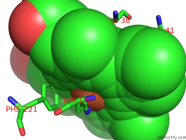
Mono view
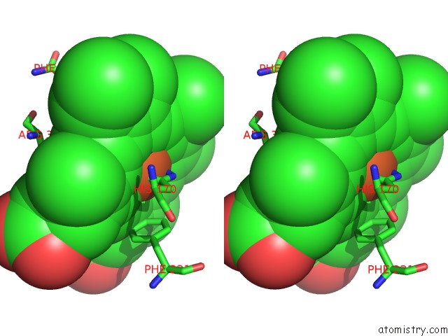
Stereo pair view
Mono view
Stereo pair view
A full contact list of Iron with other atoms in the Fe binding
site number 1 of X-Ray Induced Reduction of Horseradish Peroxidase C1A Compound III (0-100% Dose) within 5.0Å range:
|
Reference:
G.I.Berglund,
G.H.Carlsson,
A.T.Smith,
H.Szoke,
A.Henriksen,
J.Hajdu.
The Catalytic Pathway of Horseradish Peroxidase at High Resolution Nature V. 417 463 2002.
ISSN: ISSN 0028-0836
PubMed: 12024218
DOI: 10.1038/417463A
Page generated: Wed Jul 16 15:43:15 2025
ISSN: ISSN 0028-0836
PubMed: 12024218
DOI: 10.1038/417463A
Last articles
Fe in 2YXOFe in 2YRS
Fe in 2YXC
Fe in 2YNM
Fe in 2YVJ
Fe in 2YP1
Fe in 2YU2
Fe in 2YU1
Fe in 2YQB
Fe in 2YOO