Iron »
PDB 1h5g-1hdb »
1h6n »
Iron in PDB 1h6n: Formation of A Tyrosyl Radical Intermediate in Proteus Mirabilis Catalase By Directed Mutagenesis and Consequences For Nucleotide Reactivity
Enzymatic activity of Formation of A Tyrosyl Radical Intermediate in Proteus Mirabilis Catalase By Directed Mutagenesis and Consequences For Nucleotide Reactivity
All present enzymatic activity of Formation of A Tyrosyl Radical Intermediate in Proteus Mirabilis Catalase By Directed Mutagenesis and Consequences For Nucleotide Reactivity:
1.11.1.6;
1.11.1.6;
Protein crystallography data
The structure of Formation of A Tyrosyl Radical Intermediate in Proteus Mirabilis Catalase By Directed Mutagenesis and Consequences For Nucleotide Reactivity, PDB code: 1h6n
was solved by
P.Andreoletti,
G.Sainz,
M.Jaquinod,
J.Gagnon,
H.M.Jouve,
with X-Ray Crystallography technique. A brief refinement statistics is given in the table below:
| Resolution Low / High (Å) | 29.67 / 2.11 |
| Space group | P 62 2 2 |
| Cell size a, b, c (Å), α, β, γ (°) | 109.900, 109.900, 249.780, 90.00, 90.00, 120.00 |
| R / Rfree (%) | 22 / 23.6 |
Iron Binding Sites:
The binding sites of Iron atom in the Formation of A Tyrosyl Radical Intermediate in Proteus Mirabilis Catalase By Directed Mutagenesis and Consequences For Nucleotide Reactivity
(pdb code 1h6n). This binding sites where shown within
5.0 Angstroms radius around Iron atom.
In total only one binding site of Iron was determined in the Formation of A Tyrosyl Radical Intermediate in Proteus Mirabilis Catalase By Directed Mutagenesis and Consequences For Nucleotide Reactivity, PDB code: 1h6n:
In total only one binding site of Iron was determined in the Formation of A Tyrosyl Radical Intermediate in Proteus Mirabilis Catalase By Directed Mutagenesis and Consequences For Nucleotide Reactivity, PDB code: 1h6n:
Iron binding site 1 out of 1 in 1h6n
Go back to
Iron binding site 1 out
of 1 in the Formation of A Tyrosyl Radical Intermediate in Proteus Mirabilis Catalase By Directed Mutagenesis and Consequences For Nucleotide Reactivity
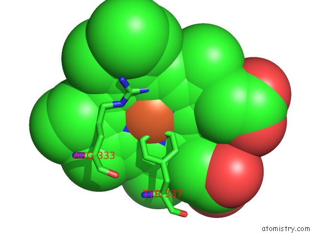
Mono view
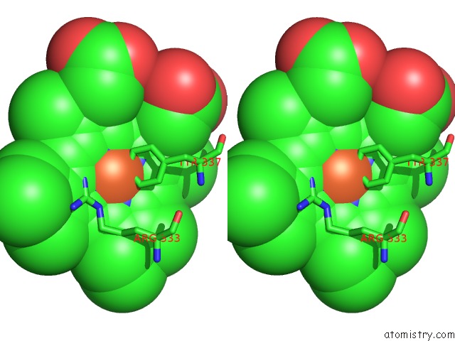
Stereo pair view
Mono view
Stereo pair view
A full contact list of Iron with other atoms in the Fe binding
site number 1 of Formation of A Tyrosyl Radical Intermediate in Proteus Mirabilis Catalase By Directed Mutagenesis and Consequences For Nucleotide Reactivity within 5.0Å range:
|
Reference:
P.Andreoletti,
G.Sainz,
M.Jaquinod,
J.Gagnon,
H.M.Jouve.
High Resolution Structure and Biochemical Properties of A Recombinant Proteus Mirabilis Catalase Depleted in Iron. Proteins: Struct.,Funct., V. 50 261 2003GENET..
ISSN: ISSN 0887-3585
PubMed: 12486720
DOI: 10.1002/PROT.10283
Page generated: Wed Jul 16 15:43:35 2025
ISSN: ISSN 0887-3585
PubMed: 12486720
DOI: 10.1002/PROT.10283
Last articles
Fe in 2YXOFe in 2YRS
Fe in 2YXC
Fe in 2YNM
Fe in 2YVJ
Fe in 2YP1
Fe in 2YU2
Fe in 2YU1
Fe in 2YQB
Fe in 2YOO