Iron »
PDB 1h5g-1hdb »
1han »
Iron in PDB 1han: Crystal Structure of the Biphenyl-Cleaving Extradiol Dioxygenase From A Pcb-Degrading Pseudomonad
Enzymatic activity of Crystal Structure of the Biphenyl-Cleaving Extradiol Dioxygenase From A Pcb-Degrading Pseudomonad
All present enzymatic activity of Crystal Structure of the Biphenyl-Cleaving Extradiol Dioxygenase From A Pcb-Degrading Pseudomonad:
1.13.11.39;
1.13.11.39;
Protein crystallography data
The structure of Crystal Structure of the Biphenyl-Cleaving Extradiol Dioxygenase From A Pcb-Degrading Pseudomonad, PDB code: 1han
was solved by
S.Han,
J.T.Bolin,
with X-Ray Crystallography technique. A brief refinement statistics is given in the table below:
| Resolution Low / High (Å) | 7.00 / 1.90 |
| Space group | I 4 2 2 |
| Cell size a, b, c (Å), α, β, γ (°) | 122.580, 122.580, 111.360, 90.00, 90.00, 90.00 |
| R / Rfree (%) | 16.2 / 19 |
Iron Binding Sites:
The binding sites of Iron atom in the Crystal Structure of the Biphenyl-Cleaving Extradiol Dioxygenase From A Pcb-Degrading Pseudomonad
(pdb code 1han). This binding sites where shown within
5.0 Angstroms radius around Iron atom.
In total 2 binding sites of Iron where determined in the Crystal Structure of the Biphenyl-Cleaving Extradiol Dioxygenase From A Pcb-Degrading Pseudomonad, PDB code: 1han:
Jump to Iron binding site number: 1; 2;
In total 2 binding sites of Iron where determined in the Crystal Structure of the Biphenyl-Cleaving Extradiol Dioxygenase From A Pcb-Degrading Pseudomonad, PDB code: 1han:
Jump to Iron binding site number: 1; 2;
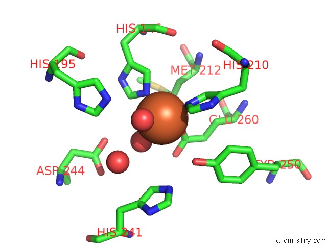
Iron binding site 1 out of 2 in 1han
Go back to
Iron binding site 1 out
of 2 in the Crystal Structure of the Biphenyl-Cleaving Extradiol Dioxygenase From A Pcb-Degrading Pseudomonad
Mono view

Stereo pair view
Mono view
Stereo pair view
A full contact list of Iron with other atoms in the Fe binding
site number 1 of Crystal Structure of the Biphenyl-Cleaving Extradiol Dioxygenase From A Pcb-Degrading Pseudomonad within 5.0Å range:
|
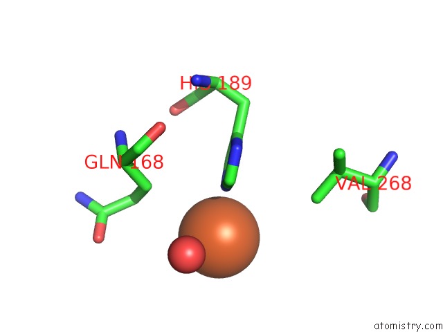
Iron binding site 2 out of 2 in 1han
Go back to
Iron binding site 2 out
of 2 in the Crystal Structure of the Biphenyl-Cleaving Extradiol Dioxygenase From A Pcb-Degrading Pseudomonad
Mono view

Stereo pair view
Mono view
Stereo pair view
A full contact list of Iron with other atoms in the Fe binding
site number 2 of Crystal Structure of the Biphenyl-Cleaving Extradiol Dioxygenase From A Pcb-Degrading Pseudomonad within 5.0Å range:
|
Reference:
S.Han,
L.D.Eltis,
K.N.Timmis,
S.W.Muchmore,
J.T.Bolin.
Crystal Structure of the Biphenyl-Cleaving Extradiol Dioxygenase From A Pcb-Degrading Pseudomonad. Science V. 270 976 1995.
ISSN: ISSN 0036-8075
PubMed: 7481800
Page generated: Sat Aug 3 07:19:32 2024
ISSN: ISSN 0036-8075
PubMed: 7481800
Last articles
Zn in 9J0NZn in 9J0O
Zn in 9J0P
Zn in 9FJX
Zn in 9EKB
Zn in 9C0F
Zn in 9CAH
Zn in 9CH0
Zn in 9CH3
Zn in 9CH1