Atomistry » Iron » PDB 1h5g-1hdb » 1hbs
Atomistry »
  Iron »
    PDB 1h5g-1hdb »
      1hbs »

Iron in PDB 1hbs: Refined Crystal Structure of Deoxyhemoglobin S. I. Restrained Least-Squares Refinement at 3.0-Angstroms Resolution

Protein crystallography data

The structure of Refined Crystal Structure of Deoxyhemoglobin S. I. Restrained Least-Squares Refinement at 3.0-Angstroms Resolution, PDB code: 1hbs was solved by E.A.Padlan, W.E.Love, with X-Ray Crystallography technique. A brief refinement statistics is given in the table below:

Resolution Low / High (Å) N/A / 3.00
Space group P 1 21 1
Cell size a, b, c (Å), α, β, γ (°) 63.330, 185.660, 52.970, 90.00, 92.69, 90.00
R / Rfree (%) 25.4 / n/a

Iron Binding Sites:

The binding sites of Iron atom in the Refined Crystal Structure of Deoxyhemoglobin S. I. Restrained Least-Squares Refinement at 3.0-Angstroms Resolution (pdb code 1hbs). This binding sites where shown within 5.0 Angstroms radius around Iron atom.
In total 8 binding sites of Iron where determined in the Refined Crystal Structure of Deoxyhemoglobin S. I. Restrained Least-Squares Refinement at 3.0-Angstroms Resolution, PDB code: 1hbs:
Jump to Iron binding site number: 1; 2; 3; 4; 5; 6; 7; 8;

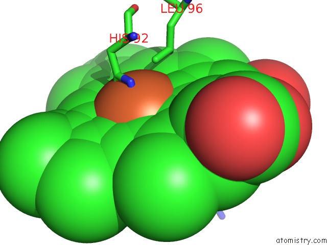
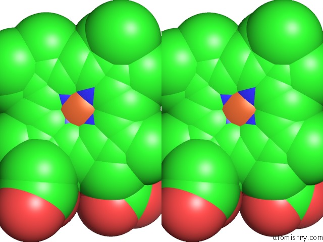
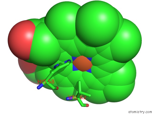
Iron binding site 1 out of 8 in 1hbs

Go back to Iron Binding Sites List in 1hbs
Iron binding site 1 out of 8 in the Refined Crystal Structure of Deoxyhemoglobin S. I. Restrained Least-Squares Refinement at 3.0-Angstroms Resolution


Mono view


Stereo pair view

A full contact list of Iron with other atoms in the Fe binding site number 1 of Refined Crystal Structure of Deoxyhemoglobin S. I. Restrained Least-Squares Refinement at 3.0-Angstroms Resolution within 5.0Å range:
probe atom residue distance (Å) B Occ
A:Fe142

b:23.5
occ:1.00
FE A:HEM142 0.0 23.5 1.0
NC A:HEM142 2.0 22.1 1.0
NB A:HEM142 2.0 20.6 1.0
ND A:HEM142 2.0 22.9 1.0
NA A:HEM142 2.2 21.6 1.0
NE2 A:HIS87 2.4 26.0 1.0
C4D A:HEM142 3.0 21.1 1.0
C4B A:HEM142 3.0 20.3 1.0
C1B A:HEM142 3.0 21.0 1.0
C4C A:HEM142 3.0 21.9 1.0
C1A A:HEM142 3.1 21.4 1.0
C4A A:HEM142 3.2 20.6 1.0
C1D A:HEM142 3.2 21.7 1.0
CE1 A:HIS87 3.2 27.7 1.0
C1C A:HEM142 3.3 21.8 1.0
CHD A:HEM142 3.3 22.7 1.0
CD2 A:HIS87 3.4 24.6 1.0
CHB A:HEM142 3.4 21.0 1.0
CHA A:HEM142 3.5 22.1 1.0
CHC A:HEM142 3.6 22.3 1.0
C2B A:HEM142 4.2 20.2 1.0
C3B A:HEM142 4.2 19.4 1.0
C3D A:HEM142 4.2 19.6 1.0
C2C A:HEM142 4.4 20.7 1.0
C3C A:HEM142 4.4 20.4 1.0
C2D A:HEM142 4.4 20.1 1.0
C2A A:HEM142 4.4 20.7 1.0
ND1 A:HIS87 4.5 25.9 1.0
CG A:HIS87 4.5 24.1 1.0
C3A A:HEM142 4.5 20.7 1.0
CZ A:PHE98 4.8 23.1 1.0

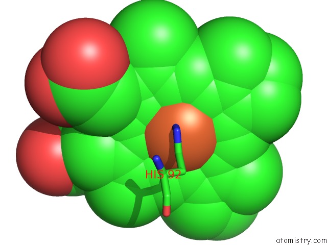
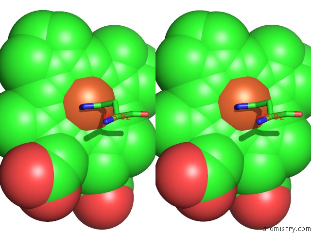
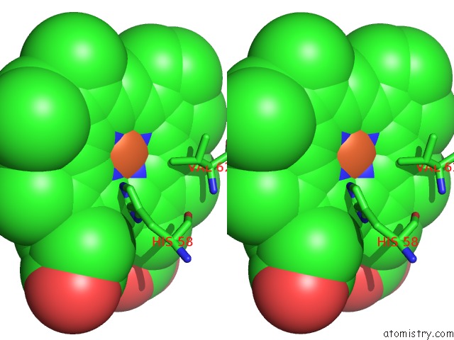
Iron binding site 2 out of 8 in 1hbs

Go back to Iron Binding Sites List in 1hbs
Iron binding site 2 out of 8 in the Refined Crystal Structure of Deoxyhemoglobin S. I. Restrained Least-Squares Refinement at 3.0-Angstroms Resolution


Mono view


Stereo pair view

A full contact list of Iron with other atoms in the Fe binding site number 2 of Refined Crystal Structure of Deoxyhemoglobin S. I. Restrained Least-Squares Refinement at 3.0-Angstroms Resolution within 5.0Å range:
probe atom residue distance (Å) B Occ
B:Fe147

b:21.8
occ:1.00
FE B:HEM147 0.0 21.8 1.0
NA B:HEM147 1.9 19.4 1.0
NE2 B:HIS92 2.0 16.0 1.0
NC B:HEM147 2.0 20.1 1.0
NB B:HEM147 2.1 17.4 1.0
ND B:HEM147 2.2 20.8 1.0
CE1 B:HIS92 2.7 14.6 1.0
CD2 B:HIS92 2.8 14.9 1.0
C4A B:HEM147 2.9 19.7 1.0
C4D B:HEM147 2.9 20.1 1.0
C1A B:HEM147 3.0 18.3 1.0
C1B B:HEM147 3.0 18.6 1.0
C4C B:HEM147 3.1 20.3 1.0
C1C B:HEM147 3.1 20.6 1.0
C4B B:HEM147 3.2 17.7 1.0
CHA B:HEM147 3.4 18.3 1.0
CHB B:HEM147 3.4 20.2 1.0
CHC B:HEM147 3.5 20.7 1.0
C1D B:HEM147 3.5 18.6 1.0
ND1 B:HIS92 3.7 14.3 1.0
CG B:HIS92 3.9 15.2 1.0
CHD B:HEM147 3.9 19.7 1.0
C2A B:HEM147 4.0 19.2 1.0
C3A B:HEM147 4.0 19.9 1.0
C2C B:HEM147 4.2 18.4 1.0
C3D B:HEM147 4.2 20.3 1.0
C2B B:HEM147 4.2 17.6 1.0
C3C B:HEM147 4.3 18.2 1.0
C3B B:HEM147 4.3 16.9 1.0
CD1 B:LEU96 4.3 24.7 1.0
C2D B:HEM147 4.5 19.9 1.0
NE2 B:HIS63 4.7 24.7 1.0
CE1 B:HIS63 4.9 24.0 1.0

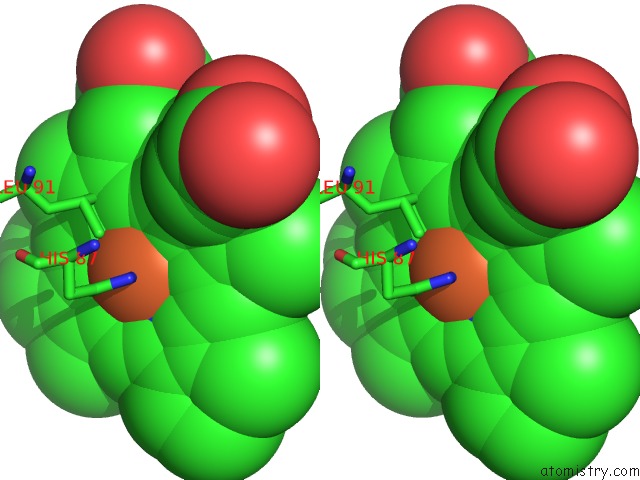
Iron binding site 3 out of 8 in 1hbs

Go back to Iron Binding Sites List in 1hbs
Iron binding site 3 out of 8 in the Refined Crystal Structure of Deoxyhemoglobin S. I. Restrained Least-Squares Refinement at 3.0-Angstroms Resolution


Mono view


Stereo pair view

A full contact list of Iron with other atoms in the Fe binding site number 3 of Refined Crystal Structure of Deoxyhemoglobin S. I. Restrained Least-Squares Refinement at 3.0-Angstroms Resolution within 5.0Å range:
probe atom residue distance (Å) B Occ
C:Fe142

b:17.3
occ:1.00
FE C:HEM142 0.0 17.3 1.0
NC C:HEM142 2.1 12.5 1.0
NA C:HEM142 2.1 16.6 1.0
NB C:HEM142 2.1 12.6 1.0
ND C:HEM142 2.1 15.1 1.0
NE2 C:HIS87 2.5 8.6 1.0
CD2 C:HIS87 2.9 10.8 1.0
C1B C:HEM142 3.0 12.4 1.0
C1A C:HEM142 3.0 18.3 1.0
C1D C:HEM142 3.1 16.4 1.0
C4C C:HEM142 3.1 13.5 1.0
C4D C:HEM142 3.1 17.3 1.0
C1C C:HEM142 3.1 11.3 1.0
C4B C:HEM142 3.1 11.4 1.0
C4A C:HEM142 3.1 16.8 1.0
CHB C:HEM142 3.3 14.5 1.0
CHA C:HEM142 3.5 18.0 1.0
CHC C:HEM142 3.5 11.0 1.0
CHD C:HEM142 3.5 15.6 1.0
CE1 C:HIS87 3.7 8.1 1.0
CG C:HIS87 4.2 11.3 1.0
C3B C:HEM142 4.3 11.1 1.0
C3C C:HEM142 4.3 12.0 1.0
C2B C:HEM142 4.3 12.1 1.0
C2C C:HEM142 4.3 10.1 1.0
C2A C:HEM142 4.3 19.3 1.0
C3D C:HEM142 4.3 18.3 1.0
C3A C:HEM142 4.3 18.0 1.0
C2D C:HEM142 4.3 18.5 1.0
ND1 C:HIS87 4.5 9.7 1.0

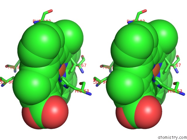
Iron binding site 4 out of 8 in 1hbs

Go back to Iron Binding Sites List in 1hbs
Iron binding site 4 out of 8 in the Refined Crystal Structure of Deoxyhemoglobin S. I. Restrained Least-Squares Refinement at 3.0-Angstroms Resolution


Mono view


Stereo pair view

A full contact list of Iron with other atoms in the Fe binding site number 4 of Refined Crystal Structure of Deoxyhemoglobin S. I. Restrained Least-Squares Refinement at 3.0-Angstroms Resolution within 5.0Å range:
probe atom residue distance (Å) B Occ
D:Fe147

b:35.2
occ:1.00
FE D:HEM147 0.0 35.2 1.0
NC D:HEM147 2.1 36.1 1.0
NE2 D:HIS92 2.1 26.8 1.0
NB D:HEM147 2.1 32.9 1.0
NA D:HEM147 2.1 33.9 1.0
ND D:HEM147 2.1 34.2 1.0
CE1 D:HIS92 2.9 26.7 1.0
C4D D:HEM147 3.0 33.1 1.0
C1B D:HEM147 3.0 33.2 1.0
C1D D:HEM147 3.1 35.5 1.0
C4B D:HEM147 3.1 32.2 1.0
C1A D:HEM147 3.1 33.1 1.0
CD2 D:HIS92 3.1 26.1 1.0
C1C D:HEM147 3.1 36.1 1.0
C4A D:HEM147 3.2 33.4 1.0
C4C D:HEM147 3.2 34.7 1.0
CHC D:HEM147 3.4 33.8 1.0
CHA D:HEM147 3.5 31.8 1.0
CHB D:HEM147 3.5 33.2 1.0
CHD D:HEM147 3.6 35.5 1.0
CG1 D:VAL67 3.6 8.7 1.0
NE2 D:HIS63 4.0 24.4 1.0
ND1 D:HIS92 4.1 27.3 1.0
CG D:HIS92 4.2 26.4 1.0
C3B D:HEM147 4.2 33.3 1.0
C2B D:HEM147 4.2 33.5 1.0
CE1 D:HIS63 4.3 24.4 1.0
C2D D:HEM147 4.3 35.3 1.0
C3D D:HEM147 4.3 34.8 1.0
C2A D:HEM147 4.3 33.2 1.0
C3C D:HEM147 4.3 34.2 1.0
C2C D:HEM147 4.4 35.9 1.0
C3A D:HEM147 4.4 33.4 1.0
CB D:VAL67 4.6 13.7 1.0
CG2 D:VAL67 4.9 11.0 1.0

Iron binding site 5 out of 8 in 1hbs

Go back to Iron Binding Sites List in 1hbs
Iron binding site 5 out of 8 in the Refined Crystal Structure of Deoxyhemoglobin S. I. Restrained Least-Squares Refinement at 3.0-Angstroms Resolution


Mono view


Stereo pair view

A full contact list of Iron with other atoms in the Fe binding site number 5 of Refined Crystal Structure of Deoxyhemoglobin S. I. Restrained Least-Squares Refinement at 3.0-Angstroms Resolution within 5.0Å range:
probe atom residue distance (Å) B Occ
E:Fe142

b:30.0
occ:1.00
FE E:HEM142 0.0 30.0 1.0
NC E:HEM142 1.9 26.9 1.0
NA E:HEM142 2.1 26.1 1.0
ND E:HEM142 2.1 24.9 1.0
NB E:HEM142 2.1 28.1 1.0
NE2 E:HIS87 2.6 21.6 1.0
C1C E:HEM142 2.9 24.0 1.0
C1D E:HEM142 2.9 24.6 1.0
C4C E:HEM142 3.0 25.4 1.0
C4A E:HEM142 3.0 26.9 1.0
C4B E:HEM142 3.0 26.5 1.0
CHD E:HEM142 3.1 24.6 1.0
C1B E:HEM142 3.1 28.5 1.0
CD2 E:HIS87 3.2 19.5 1.0
C1A E:HEM142 3.2 27.3 1.0
C4D E:HEM142 3.2 26.4 1.0
CHC E:HEM142 3.3 24.6 1.0
CHB E:HEM142 3.6 27.6 1.0
CHA E:HEM142 3.6 28.1 1.0
CE1 E:HIS87 3.7 23.0 1.0
C2C E:HEM142 4.0 22.6 1.0
C3C E:HEM142 4.1 23.9 1.0
NE2 E:HIS58 4.2 9.6 1.0
C3B E:HEM142 4.2 27.6 1.0
C2D E:HEM142 4.2 24.5 1.0
C2B E:HEM142 4.3 27.2 1.0
C3D E:HEM142 4.3 25.6 1.0
C3A E:HEM142 4.4 27.5 1.0
CG E:HIS87 4.4 20.1 1.0
C2A E:HEM142 4.5 27.0 1.0
ND1 E:HIS87 4.7 22.8 1.0
CE1 E:HIS58 4.8 6.8 1.0
CG2 E:VAL62 5.0 20.5 1.0

Iron binding site 6 out of 8 in 1hbs

Go back to Iron Binding Sites List in 1hbs
Iron binding site 6 out of 8 in the Refined Crystal Structure of Deoxyhemoglobin S. I. Restrained Least-Squares Refinement at 3.0-Angstroms Resolution


Mono view


Stereo pair view

A full contact list of Iron with other atoms in the Fe binding site number 6 of Refined Crystal Structure of Deoxyhemoglobin S. I. Restrained Least-Squares Refinement at 3.0-Angstroms Resolution within 5.0Å range:
probe atom residue distance (Å) B Occ
F:Fe147

b:25.9
occ:1.00
FE F:HEM147 0.0 25.9 1.0
NB F:HEM147 1.9 25.5 1.0
ND F:HEM147 2.0 26.7 1.0
NC F:HEM147 2.0 24.5 1.0
NA F:HEM147 2.2 29.3 1.0
NE2 F:HIS92 2.6 22.6 1.0
C4B F:HEM147 2.8 24.7 1.0
C4C F:HEM147 2.9 24.6 1.0
C4D F:HEM147 3.0 27.4 1.0
C1B F:HEM147 3.0 25.8 1.0
C1A F:HEM147 3.1 29.2 1.0
C1D F:HEM147 3.1 26.4 1.0
C1C F:HEM147 3.1 24.0 1.0
CE1 F:HIS92 3.2 22.8 1.0
CHA F:HEM147 3.4 28.3 1.0
CG2 F:VAL67 3.4 18.3 1.0
CHB F:HEM147 3.4 28.9 1.0
C4A F:HEM147 3.4 29.6 1.0
CHD F:HEM147 3.5 26.9 1.0
CHC F:HEM147 3.6 23.3 1.0
CD2 F:HIS92 3.7 21.8 1.0
CE1 F:HIS63 3.8 21.9 1.0
C3B F:HEM147 4.1 24.1 1.0
C2B F:HEM147 4.1 24.2 1.0
C3D F:HEM147 4.3 27.7 1.0
C2D F:HEM147 4.3 24.9 1.0
NE2 F:HIS63 4.3 22.0 1.0
C3C F:HEM147 4.3 21.8 1.0
C2C F:HEM147 4.3 22.3 1.0
ND1 F:HIS92 4.4 21.4 1.0
C2A F:HEM147 4.5 30.5 1.0
C3A F:HEM147 4.5 31.1 1.0
ND1 F:HIS63 4.6 23.3 1.0
CB F:VAL67 4.7 16.9 1.0
CG F:HIS92 4.8 21.1 1.0

Iron binding site 7 out of 8 in 1hbs

Go back to Iron Binding Sites List in 1hbs
Iron binding site 7 out of 8 in the Refined Crystal Structure of Deoxyhemoglobin S. I. Restrained Least-Squares Refinement at 3.0-Angstroms Resolution


Mono view


Stereo pair view

A full contact list of Iron with other atoms in the Fe binding site number 7 of Refined Crystal Structure of Deoxyhemoglobin S. I. Restrained Least-Squares Refinement at 3.0-Angstroms Resolution within 5.0Å range:
probe atom residue distance (Å) B Occ
G:Fe142

b:22.7
occ:1.00
FE G:HEM142 0.0 22.7 1.0
NC G:HEM142 1.9 24.9 1.0
NA G:HEM142 1.9 23.0 1.0
ND G:HEM142 2.1 24.1 1.0
NB G:HEM142 2.3 23.2 1.0
NE2 G:HIS87 2.7 4.7 1.0
CE1 G:HIS87 2.7 4.4 1.0
C1A G:HEM142 2.8 22.1 1.0
C4C G:HEM142 3.0 26.3 1.0
C1C G:HEM142 3.0 24.7 1.0
C4D G:HEM142 3.0 25.7 1.0
C1D G:HEM142 3.0 24.6 1.0
C4A G:HEM142 3.1 24.6 1.0
CHA G:HEM142 3.2 23.1 1.0
C4B G:HEM142 3.3 22.7 1.0
CHD G:HEM142 3.3 24.5 1.0
C1B G:HEM142 3.4 22.2 1.0
CHC G:HEM142 3.5 24.4 1.0
CE1 G:HIS58 3.6 42.1 1.0
CHB G:HEM142 3.8 22.8 1.0
CD2 G:HIS87 4.0 3.5 1.0
ND1 G:HIS87 4.0 4.4 1.0
C2A G:HEM142 4.1 23.6 1.0
C2C G:HEM142 4.2 25.2 1.0
C3C G:HEM142 4.2 27.0 1.0
C3A G:HEM142 4.3 24.2 1.0
C3D G:HEM142 4.3 27.8 1.0
C2D G:HEM142 4.4 26.2 1.0
ND1 G:HIS58 4.4 40.7 1.0
CD2 G:LEU91 4.4 19.0 1.0
NE2 G:HIS58 4.4 42.0 1.0
C3B G:HEM142 4.5 21.5 1.0
CD1 G:LEU91 4.5 17.8 1.0
C2B G:HEM142 4.6 22.3 1.0
CG G:HIS87 4.7 6.6 1.0

Iron binding site 8 out of 8 in 1hbs

Go back to Iron Binding Sites List in 1hbs
Iron binding site 8 out of 8 in the Refined Crystal Structure of Deoxyhemoglobin S. I. Restrained Least-Squares Refinement at 3.0-Angstroms Resolution


Mono view


Stereo pair view

A full contact list of Iron with other atoms in the Fe binding site number 8 of Refined Crystal Structure of Deoxyhemoglobin S. I. Restrained Least-Squares Refinement at 3.0-Angstroms Resolution within 5.0Å range:
probe atom residue distance (Å) B Occ
H:Fe147

b:26.8
occ:1.00
FE H:HEM147 0.0 26.8 1.0
NB H:HEM147 2.0 23.9 1.0
NA H:HEM147 2.1 25.6 1.0
NC H:HEM147 2.1 23.4 1.0
ND H:HEM147 2.1 24.3 1.0
CE1 H:HIS92 2.4 14.9 1.0
NE2 H:HIS92 2.8 17.8 1.0
C1C H:HEM147 3.0 21.6 1.0
C1D H:HEM147 3.0 24.6 1.0
C1B H:HEM147 3.0 23.3 1.0
C4A H:HEM147 3.0 24.5 1.0
C4B H:HEM147 3.1 22.5 1.0
C1A H:HEM147 3.1 25.8 1.0
C4D H:HEM147 3.2 24.4 1.0
C4C H:HEM147 3.2 23.7 1.0
CHC H:HEM147 3.2 20.9 1.0
CHB H:HEM147 3.4 23.5 1.0
CHD H:HEM147 3.5 24.8 1.0
CHA H:HEM147 3.5 24.1 1.0
ND1 H:HIS92 3.6 12.4 1.0
CD2 H:HIS92 4.1 15.5 1.0
C2C H:HEM147 4.1 22.7 1.0
C2B H:HEM147 4.2 22.5 1.0
C3B H:HEM147 4.2 22.8 1.0
C3A H:HEM147 4.2 24.9 1.0
C3C H:HEM147 4.3 22.9 1.0
C2A H:HEM147 4.3 26.3 1.0
C2D H:HEM147 4.3 22.9 1.0
NE2 H:HIS63 4.4 16.2 1.0
CG H:HIS92 4.4 14.4 1.0
C3D H:HEM147 4.4 23.3 1.0
CE1 H:PHE103 4.5 15.8 1.0
CG1 H:VAL67 4.7 20.4 1.0
CG2 H:VAL67 4.7 19.8 1.0
CE1 H:HIS63 4.7 14.3 1.0
CD1 H:LEU96 4.7 11.4 1.0

Reference:

E.A.Padlan, W.E.Love. Refined Crystal Structure of Deoxyhemoglobin S. I. Restrained Least-Squares Refinement at 3.0-A Resolution. J.Biol.Chem. V. 260 8272 1985.
ISSN: ISSN 0021-9258
PubMed: 4008491
Page generated: Sun Dec 13 14:17:27 2020

Last articles

Zn in 8WB0
Zn in 8WAX
Zn in 8WAU
Zn in 8WAZ
Zn in 8WAY
Zn in 8WAV
Zn in 8WAW
Zn in 8WAT
Zn in 8W7M
Zn in 8WD3
© Copyright 2008-2020 by atomistry.com
Home   |    Site Map   |    Copyright   |    Contact us   |    Privacy