Iron »
PDB 1h5g-1hdb »
1hdb »
Iron in PDB 1hdb: Analysis of the Crystal Structure, Molecular Modeling and Infrared Spectroscopy of the Distal Beta-Heme Pocket VALINE67(E11)-Threonine Mutation of Hemoglobin
Protein crystallography data
The structure of Analysis of the Crystal Structure, Molecular Modeling and Infrared Spectroscopy of the Distal Beta-Heme Pocket VALINE67(E11)-Threonine Mutation of Hemoglobin, PDB code: 1hdb
was solved by
I.Pechik,
X.Ji,
C.Fronticelli,
G.L.Gilliland,
with X-Ray Crystallography technique. A brief refinement statistics is given in the table below:
| Resolution Low / High (Å) | 6.00 / 2.20 |
| Space group | P 1 21 1 |
| Cell size a, b, c (Å), α, β, γ (°) | 63.540, 83.190, 54.020, 90.00, 99.15, 90.00 |
| R / Rfree (%) | n/a / n/a |
Iron Binding Sites:
The binding sites of Iron atom in the Analysis of the Crystal Structure, Molecular Modeling and Infrared Spectroscopy of the Distal Beta-Heme Pocket VALINE67(E11)-Threonine Mutation of Hemoglobin
(pdb code 1hdb). This binding sites where shown within
5.0 Angstroms radius around Iron atom.
In total 4 binding sites of Iron where determined in the Analysis of the Crystal Structure, Molecular Modeling and Infrared Spectroscopy of the Distal Beta-Heme Pocket VALINE67(E11)-Threonine Mutation of Hemoglobin, PDB code: 1hdb:
Jump to Iron binding site number: 1; 2; 3; 4;
In total 4 binding sites of Iron where determined in the Analysis of the Crystal Structure, Molecular Modeling and Infrared Spectroscopy of the Distal Beta-Heme Pocket VALINE67(E11)-Threonine Mutation of Hemoglobin, PDB code: 1hdb:
Jump to Iron binding site number: 1; 2; 3; 4;
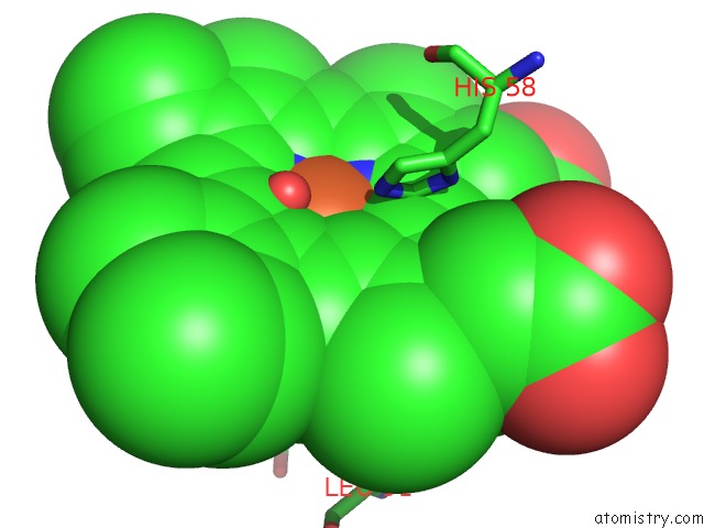
Iron binding site 1 out of 4 in 1hdb
Go back to
Iron binding site 1 out
of 4 in the Analysis of the Crystal Structure, Molecular Modeling and Infrared Spectroscopy of the Distal Beta-Heme Pocket VALINE67(E11)-Threonine Mutation of Hemoglobin
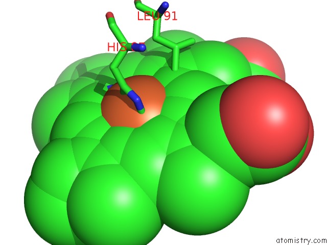
Mono view
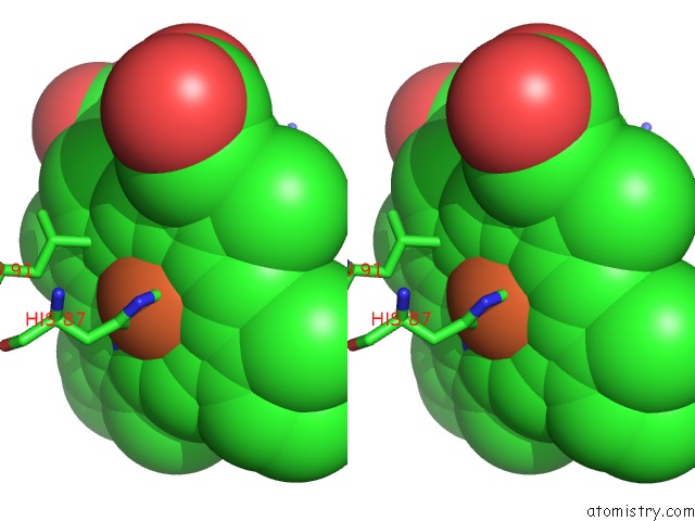
Stereo pair view
Mono view
Stereo pair view
A full contact list of Iron with other atoms in the Fe binding
site number 1 of Analysis of the Crystal Structure, Molecular Modeling and Infrared Spectroscopy of the Distal Beta-Heme Pocket VALINE67(E11)-Threonine Mutation of Hemoglobin within 5.0Å range:
|
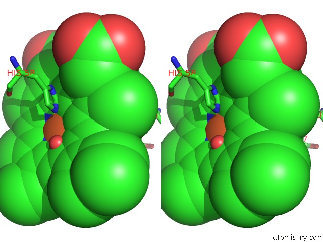
Iron binding site 2 out of 4 in 1hdb
Go back to
Iron binding site 2 out
of 4 in the Analysis of the Crystal Structure, Molecular Modeling and Infrared Spectroscopy of the Distal Beta-Heme Pocket VALINE67(E11)-Threonine Mutation of Hemoglobin
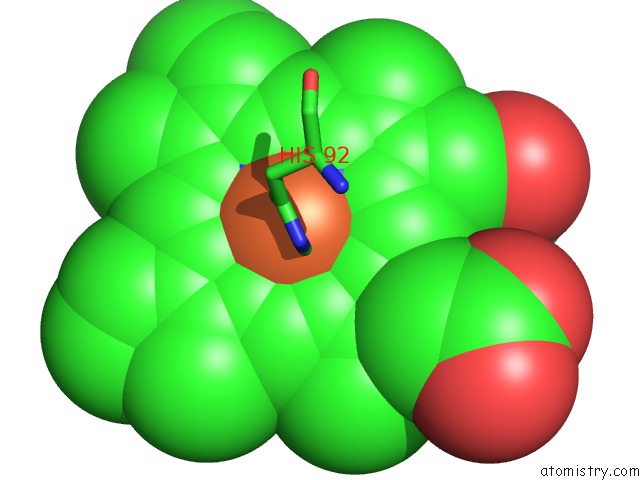
Mono view
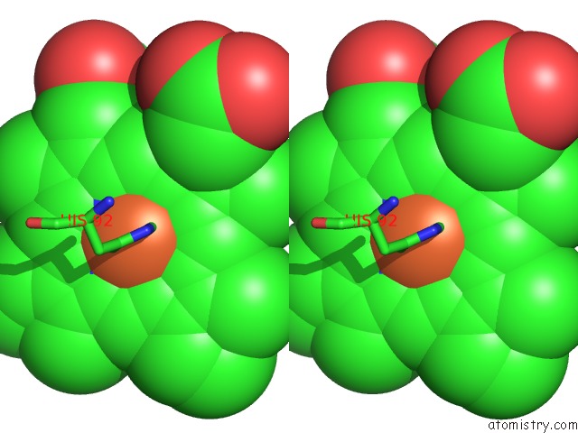
Stereo pair view
Mono view
Stereo pair view
A full contact list of Iron with other atoms in the Fe binding
site number 2 of Analysis of the Crystal Structure, Molecular Modeling and Infrared Spectroscopy of the Distal Beta-Heme Pocket VALINE67(E11)-Threonine Mutation of Hemoglobin within 5.0Å range:
|
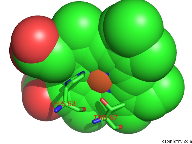
Iron binding site 3 out of 4 in 1hdb
Go back to
Iron binding site 3 out
of 4 in the Analysis of the Crystal Structure, Molecular Modeling and Infrared Spectroscopy of the Distal Beta-Heme Pocket VALINE67(E11)-Threonine Mutation of Hemoglobin
Mono view
Stereo pair view
Mono view
Stereo pair view
A full contact list of Iron with other atoms in the Fe binding
site number 3 of Analysis of the Crystal Structure, Molecular Modeling and Infrared Spectroscopy of the Distal Beta-Heme Pocket VALINE67(E11)-Threonine Mutation of Hemoglobin within 5.0Å range:
|
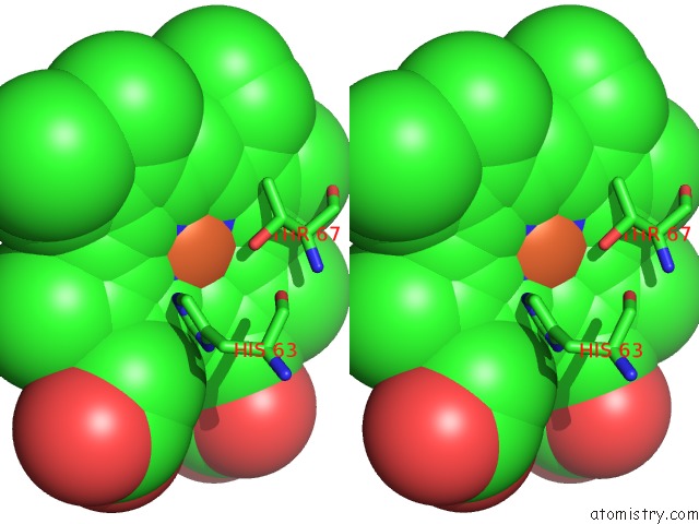
Iron binding site 4 out of 4 in 1hdb
Go back to
Iron binding site 4 out
of 4 in the Analysis of the Crystal Structure, Molecular Modeling and Infrared Spectroscopy of the Distal Beta-Heme Pocket VALINE67(E11)-Threonine Mutation of Hemoglobin
Mono view
Stereo pair view
Mono view
Stereo pair view
A full contact list of Iron with other atoms in the Fe binding
site number 4 of Analysis of the Crystal Structure, Molecular Modeling and Infrared Spectroscopy of the Distal Beta-Heme Pocket VALINE67(E11)-Threonine Mutation of Hemoglobin within 5.0Å range:
|
Reference:
I.Pechik,
X.Ji,
K.Fidelis,
M.Karavitis,
J.Moult,
W.S.Brinigar,
C.Fronticelli,
G.L.Gilliland.
Crystallographic, Molecular Modeling, and Biophysical Characterization of the Valine Beta 67 (E11)-->Threonine Variant of Hemoglobin. Biochemistry V. 35 1935 1996.
ISSN: ISSN 0006-2960
PubMed: 8639677
DOI: 10.1021/BI9519967
Page generated: Sat Aug 3 07:26:15 2024
ISSN: ISSN 0006-2960
PubMed: 8639677
DOI: 10.1021/BI9519967
Last articles
Zn in 9J0NZn in 9J0O
Zn in 9J0P
Zn in 9FJX
Zn in 9EKB
Zn in 9C0F
Zn in 9CAH
Zn in 9CH0
Zn in 9CH3
Zn in 9CH1