Iron »
PDB 1hds-1hxq »
1hlq »
Iron in PDB 1hlq: Crystal Structure of Rhodoferax Fermentans High Potential Iron-Sulfur Protein Refined to 1.45 A
Protein crystallography data
The structure of Crystal Structure of Rhodoferax Fermentans High Potential Iron-Sulfur Protein Refined to 1.45 A, PDB code: 1hlq
was solved by
A.Gonzalez,
S.Ciurli,
S.Benini,
with X-Ray Crystallography technique. A brief refinement statistics is given in the table below:
| Resolution Low / High (Å) | 22.00 / 1.45 |
| Space group | P 43 21 2 |
| Cell size a, b, c (Å), α, β, γ (°) | 88.312, 88.312, 61.340, 90.00, 90.00, 90.00 |
| R / Rfree (%) | 18.7 / 21.8 |
Iron Binding Sites:
Pages:
>>> Page 1 <<< Page 2, Binding sites: 11 - 12;Binding sites:
The binding sites of Iron atom in the Crystal Structure of Rhodoferax Fermentans High Potential Iron-Sulfur Protein Refined to 1.45 A (pdb code 1hlq). This binding sites where shown within 5.0 Angstroms radius around Iron atom.In total 12 binding sites of Iron where determined in the Crystal Structure of Rhodoferax Fermentans High Potential Iron-Sulfur Protein Refined to 1.45 A, PDB code: 1hlq:
Jump to Iron binding site number: 1; 2; 3; 4; 5; 6; 7; 8; 9; 10;
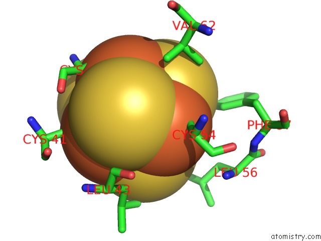
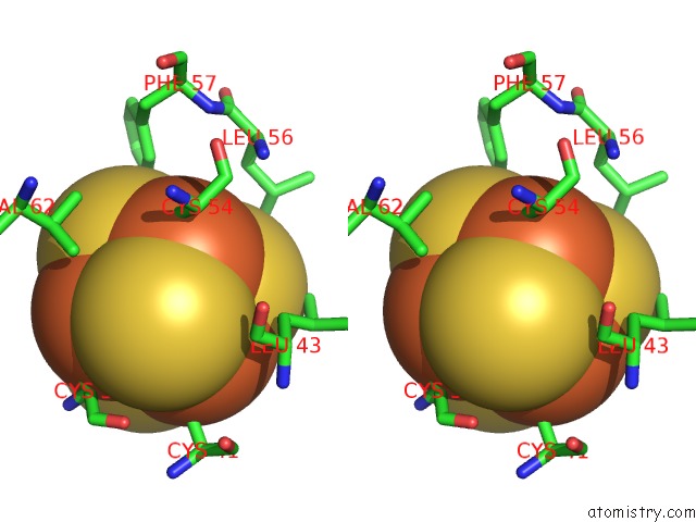
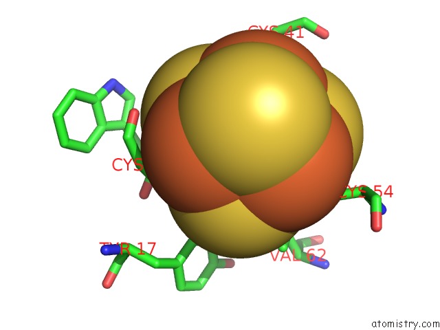
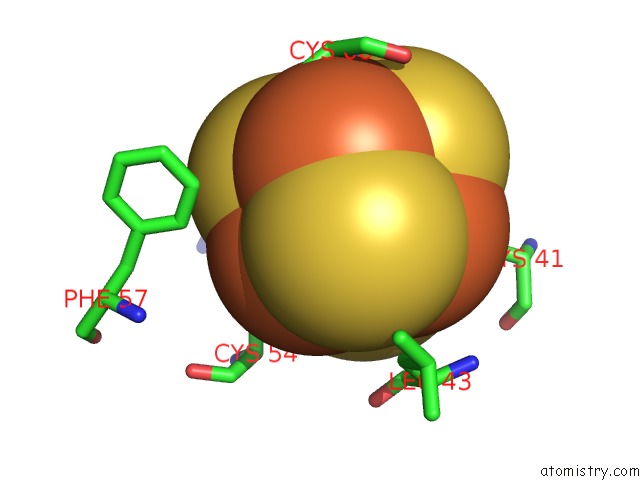
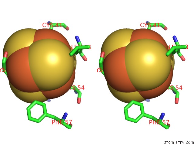
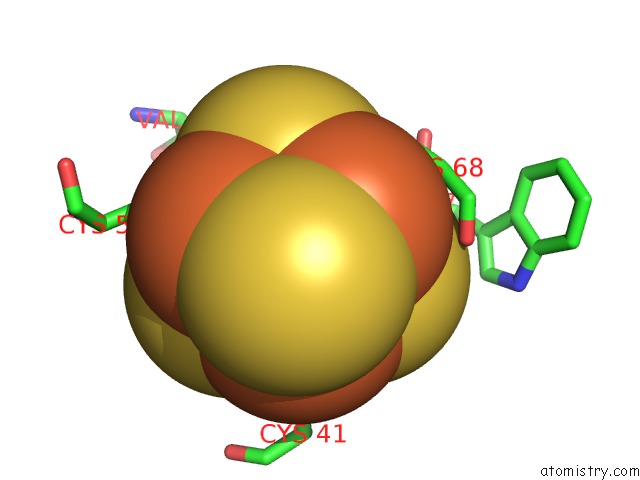
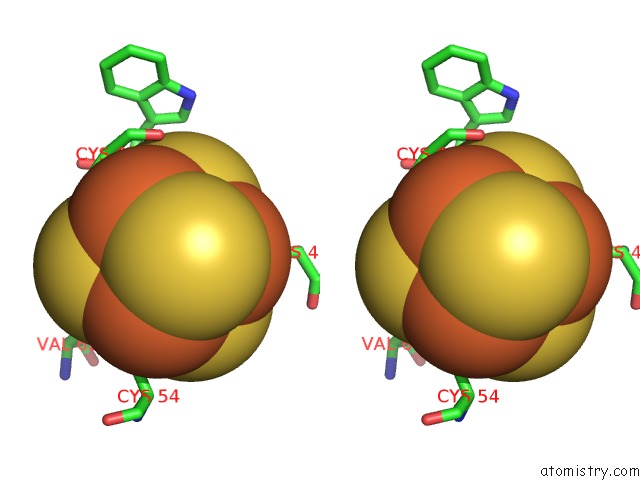
Iron binding site 1 out of 12 in 1hlq
Go back to
Iron binding site 1 out
of 12 in the Crystal Structure of Rhodoferax Fermentans High Potential Iron-Sulfur Protein Refined to 1.45 A
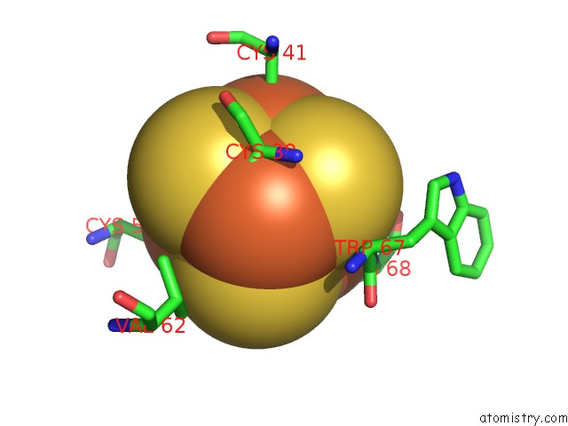
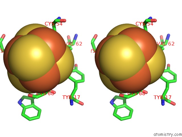
Mono view
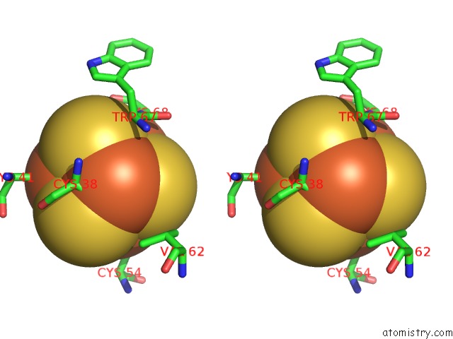
Stereo pair view
Mono view
Stereo pair view
A full contact list of Iron with other atoms in the Fe binding
site number 1 of Crystal Structure of Rhodoferax Fermentans High Potential Iron-Sulfur Protein Refined to 1.45 A within 5.0Å range:
|
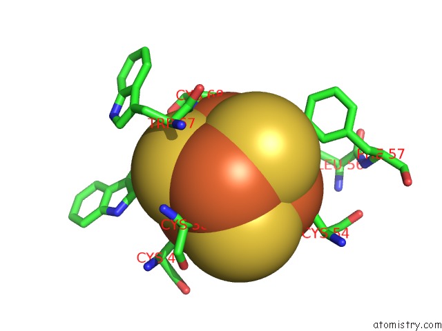
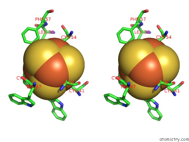
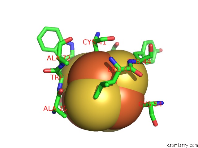
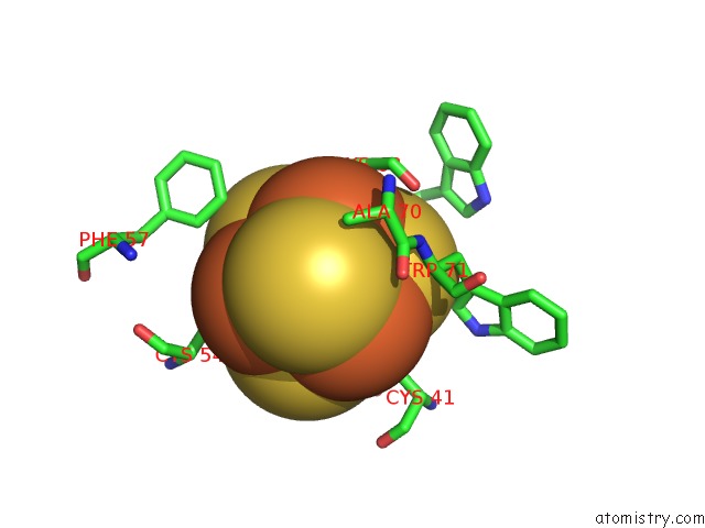
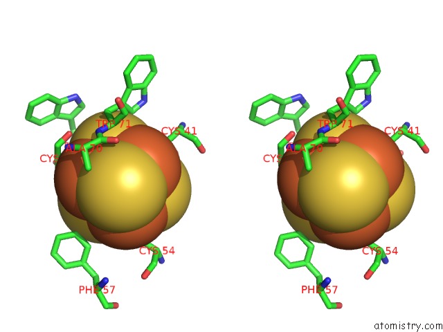
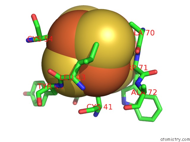
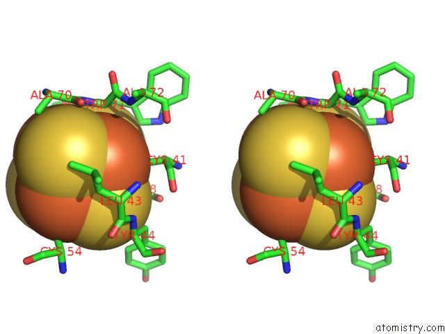
Iron binding site 2 out of 12 in 1hlq
Go back to
Iron binding site 2 out
of 12 in the Crystal Structure of Rhodoferax Fermentans High Potential Iron-Sulfur Protein Refined to 1.45 A
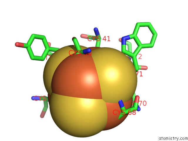
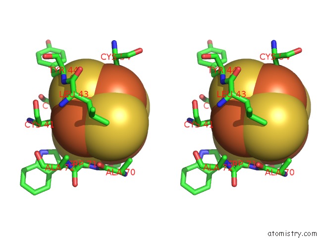
Mono view
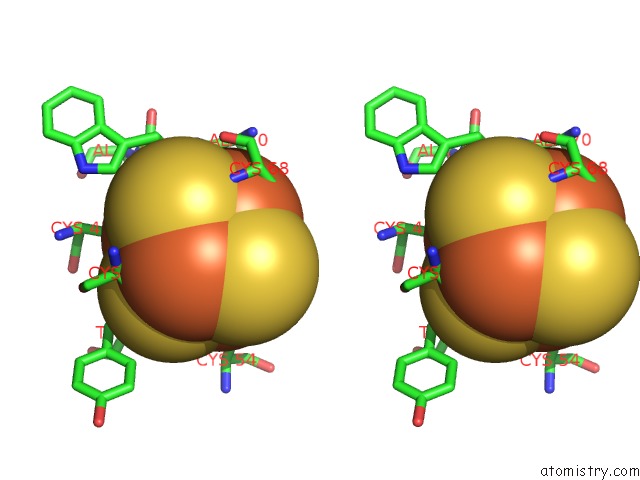
Stereo pair view
Mono view
Stereo pair view
A full contact list of Iron with other atoms in the Fe binding
site number 2 of Crystal Structure of Rhodoferax Fermentans High Potential Iron-Sulfur Protein Refined to 1.45 A within 5.0Å range:
|
Iron binding site 3 out of 12 in 1hlq
Go back to
Iron binding site 3 out
of 12 in the Crystal Structure of Rhodoferax Fermentans High Potential Iron-Sulfur Protein Refined to 1.45 A
Mono view
Stereo pair view
Mono view
Stereo pair view
A full contact list of Iron with other atoms in the Fe binding
site number 3 of Crystal Structure of Rhodoferax Fermentans High Potential Iron-Sulfur Protein Refined to 1.45 A within 5.0Å range:
|
Iron binding site 4 out of 12 in 1hlq
Go back to
Iron binding site 4 out
of 12 in the Crystal Structure of Rhodoferax Fermentans High Potential Iron-Sulfur Protein Refined to 1.45 A
Mono view
Stereo pair view
Mono view
Stereo pair view
A full contact list of Iron with other atoms in the Fe binding
site number 4 of Crystal Structure of Rhodoferax Fermentans High Potential Iron-Sulfur Protein Refined to 1.45 A within 5.0Å range:
|
Iron binding site 5 out of 12 in 1hlq
Go back to
Iron binding site 5 out
of 12 in the Crystal Structure of Rhodoferax Fermentans High Potential Iron-Sulfur Protein Refined to 1.45 A
Mono view
Stereo pair view
Mono view
Stereo pair view
A full contact list of Iron with other atoms in the Fe binding
site number 5 of Crystal Structure of Rhodoferax Fermentans High Potential Iron-Sulfur Protein Refined to 1.45 A within 5.0Å range:
|
Iron binding site 6 out of 12 in 1hlq
Go back to
Iron binding site 6 out
of 12 in the Crystal Structure of Rhodoferax Fermentans High Potential Iron-Sulfur Protein Refined to 1.45 A
Mono view
Stereo pair view
Mono view
Stereo pair view
A full contact list of Iron with other atoms in the Fe binding
site number 6 of Crystal Structure of Rhodoferax Fermentans High Potential Iron-Sulfur Protein Refined to 1.45 A within 5.0Å range:
|
Iron binding site 7 out of 12 in 1hlq
Go back to
Iron binding site 7 out
of 12 in the Crystal Structure of Rhodoferax Fermentans High Potential Iron-Sulfur Protein Refined to 1.45 A
Mono view
Stereo pair view
Mono view
Stereo pair view
A full contact list of Iron with other atoms in the Fe binding
site number 7 of Crystal Structure of Rhodoferax Fermentans High Potential Iron-Sulfur Protein Refined to 1.45 A within 5.0Å range:
|
Iron binding site 8 out of 12 in 1hlq
Go back to
Iron binding site 8 out
of 12 in the Crystal Structure of Rhodoferax Fermentans High Potential Iron-Sulfur Protein Refined to 1.45 A
Mono view
Stereo pair view
Mono view
Stereo pair view
A full contact list of Iron with other atoms in the Fe binding
site number 8 of Crystal Structure of Rhodoferax Fermentans High Potential Iron-Sulfur Protein Refined to 1.45 A within 5.0Å range:
|
Iron binding site 9 out of 12 in 1hlq
Go back to
Iron binding site 9 out
of 12 in the Crystal Structure of Rhodoferax Fermentans High Potential Iron-Sulfur Protein Refined to 1.45 A
Mono view
Stereo pair view
Mono view
Stereo pair view
A full contact list of Iron with other atoms in the Fe binding
site number 9 of Crystal Structure of Rhodoferax Fermentans High Potential Iron-Sulfur Protein Refined to 1.45 A within 5.0Å range:
|
Iron binding site 10 out of 12 in 1hlq
Go back to
Iron binding site 10 out
of 12 in the Crystal Structure of Rhodoferax Fermentans High Potential Iron-Sulfur Protein Refined to 1.45 A
Mono view
Stereo pair view
Mono view
Stereo pair view
A full contact list of Iron with other atoms in the Fe binding
site number 10 of Crystal Structure of Rhodoferax Fermentans High Potential Iron-Sulfur Protein Refined to 1.45 A within 5.0Å range:
|
Reference:
A.Gonzalez,
S.Benini,
S.Ciurli.
Structure of Rhodoferax Fermentans High-Potential Iron-Sulfur Protein Solved By Mad. Acta Crystallogr.,Sect.D V. 59 1582 2003.
ISSN: ISSN 0907-4449
PubMed: 12925788
DOI: 10.1107/S0907444903014604
Page generated: Sat Aug 3 07:46:32 2024
ISSN: ISSN 0907-4449
PubMed: 12925788
DOI: 10.1107/S0907444903014604
Last articles
Zn in 9J0NZn in 9J0O
Zn in 9J0P
Zn in 9FJX
Zn in 9EKB
Zn in 9C0F
Zn in 9CAH
Zn in 9CH0
Zn in 9CH3
Zn in 9CH1