Iron »
PDB 1hds-1hxq »
1hse »
Iron in PDB 1hse: H253M N Terminal Lobe of Human Lactoferrin
Protein crystallography data
The structure of H253M N Terminal Lobe of Human Lactoferrin, PDB code: 1hse
was solved by
H.Nicholson,
B.F.Anderson,
E.N.Baker,
with X-Ray Crystallography technique. A brief refinement statistics is given in the table below:
| Resolution Low / High (Å) | 20.00 / 2.20 |
| Space group | P 41 21 2 |
| Cell size a, b, c (Å), α, β, γ (°) | 58.400, 58.400, 217.900, 90.00, 90.00, 90.00 |
| R / Rfree (%) | 17.8 / n/a |
Iron Binding Sites:
The binding sites of Iron atom in the H253M N Terminal Lobe of Human Lactoferrin
(pdb code 1hse). This binding sites where shown within
5.0 Angstroms radius around Iron atom.
In total only one binding site of Iron was determined in the H253M N Terminal Lobe of Human Lactoferrin, PDB code: 1hse:
In total only one binding site of Iron was determined in the H253M N Terminal Lobe of Human Lactoferrin, PDB code: 1hse:
Iron binding site 1 out of 1 in 1hse
Go back to
Iron binding site 1 out
of 1 in the H253M N Terminal Lobe of Human Lactoferrin

Mono view
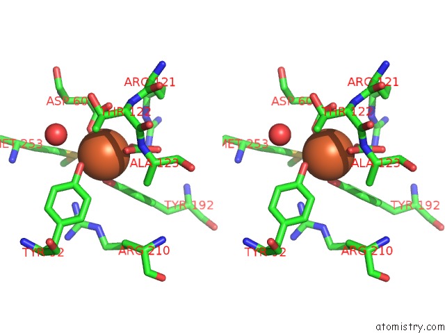
Stereo pair view
Mono view
Stereo pair view
A full contact list of Iron with other atoms in the Fe binding
site number 1 of H253M N Terminal Lobe of Human Lactoferrin within 5.0Å range:
|
Reference:
H.Nicholson,
B.F.Anderson,
T.Bland,
S.C.Shewry,
J.W.Tweedie,
E.N.Baker.
Mutagenesis of the Histidine Ligand in Human Lactoferrin: Iron Binding Properties and Crystal Structure of the Histidine-253-->Methionine Mutant. Biochemistry V. 36 341 1997.
ISSN: ISSN 0006-2960
PubMed: 9003186
DOI: 10.1021/BI961908Y
Page generated: Sat Aug 3 07:49:07 2024
ISSN: ISSN 0006-2960
PubMed: 9003186
DOI: 10.1021/BI961908Y
Last articles
Cl in 5SYMCl in 5SYV
Cl in 5SYU
Cl in 5SYL
Cl in 5SYK
Cl in 5SYJ
Cl in 5SYI
Cl in 5SYH
Cl in 5SXX
Cl in 5SXT