Iron »
PDB 1hds-1hxq »
1ht8 »
Iron in PDB 1ht8: The 2.7 Angstrom Resolution Model of Ovine Cox-1 Complexed with Alclofenac
Enzymatic activity of The 2.7 Angstrom Resolution Model of Ovine Cox-1 Complexed with Alclofenac
All present enzymatic activity of The 2.7 Angstrom Resolution Model of Ovine Cox-1 Complexed with Alclofenac:
1.14.99.1;
1.14.99.1;
Protein crystallography data
The structure of The 2.7 Angstrom Resolution Model of Ovine Cox-1 Complexed with Alclofenac, PDB code: 1ht8
was solved by
B.S.Selinsky,
K.Gupta,
C.T.Sharkey,
P.J.Loll,
with X-Ray Crystallography technique. A brief refinement statistics is given in the table below:
| Resolution Low / High (Å) | 19.92 / 2.69 |
| Space group | I 2 2 2 |
| Cell size a, b, c (Å), α, β, γ (°) | 99.149, 208.450, 222.399, 90.00, 90.00, 90.00 |
| R / Rfree (%) | 21.1 / 24.2 |
Other elements in 1ht8:
The structure of The 2.7 Angstrom Resolution Model of Ovine Cox-1 Complexed with Alclofenac also contains other interesting chemical elements:
| Chlorine | (Cl) | 2 atoms |
Iron Binding Sites:
The binding sites of Iron atom in the The 2.7 Angstrom Resolution Model of Ovine Cox-1 Complexed with Alclofenac
(pdb code 1ht8). This binding sites where shown within
5.0 Angstroms radius around Iron atom.
In total 2 binding sites of Iron where determined in the The 2.7 Angstrom Resolution Model of Ovine Cox-1 Complexed with Alclofenac, PDB code: 1ht8:
Jump to Iron binding site number: 1; 2;
In total 2 binding sites of Iron where determined in the The 2.7 Angstrom Resolution Model of Ovine Cox-1 Complexed with Alclofenac, PDB code: 1ht8:
Jump to Iron binding site number: 1; 2;
Iron binding site 1 out of 2 in 1ht8
Go back to
Iron binding site 1 out
of 2 in the The 2.7 Angstrom Resolution Model of Ovine Cox-1 Complexed with Alclofenac
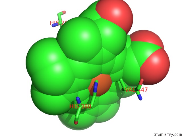
Mono view
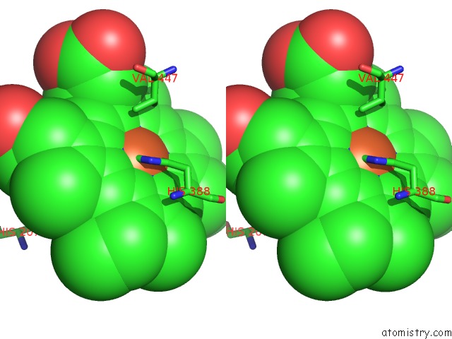
Stereo pair view
Mono view
Stereo pair view
A full contact list of Iron with other atoms in the Fe binding
site number 1 of The 2.7 Angstrom Resolution Model of Ovine Cox-1 Complexed with Alclofenac within 5.0Å range:
|
Iron binding site 2 out of 2 in 1ht8
Go back to
Iron binding site 2 out
of 2 in the The 2.7 Angstrom Resolution Model of Ovine Cox-1 Complexed with Alclofenac
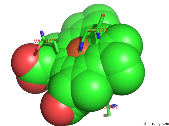
Mono view
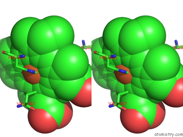
Stereo pair view
Mono view
Stereo pair view
A full contact list of Iron with other atoms in the Fe binding
site number 2 of The 2.7 Angstrom Resolution Model of Ovine Cox-1 Complexed with Alclofenac within 5.0Å range:
|
Reference:
B.S.Selinsky,
K.Gupta,
C.T.Sharkey,
P.J.Loll.
Structural Analysis of Nsaid Binding By Prostaglandin H2 Synthase: Time-Dependent and Time-Independent Inhibitors Elicit Identical Enzyme Conformations. Biochemistry V. 40 5172 2001.
ISSN: ISSN 0006-2960
PubMed: 11318639
DOI: 10.1021/BI010045S
Page generated: Wed Jul 16 16:07:53 2025
ISSN: ISSN 0006-2960
PubMed: 11318639
DOI: 10.1021/BI010045S
Last articles
Fe in 2YXOFe in 2YRS
Fe in 2YXC
Fe in 2YNM
Fe in 2YVJ
Fe in 2YP1
Fe in 2YU2
Fe in 2YU1
Fe in 2YQB
Fe in 2YOO