Iron »
PDB 1hds-1hxq »
1hv4 »
Iron in PDB 1hv4: Crystal Structure Analysis of Bar-Head Goose Hemoglobin (Deoxy Form)
Protein crystallography data
The structure of Crystal Structure Analysis of Bar-Head Goose Hemoglobin (Deoxy Form), PDB code: 1hv4
was solved by
Y.Liang,
Z.Hua,
X.Liang,
Q.Xu,
G.Lu,
with X-Ray Crystallography technique. A brief refinement statistics is given in the table below:
| Resolution Low / High (Å) | 36.89 / 2.80 |
| Space group | P 1 |
| Cell size a, b, c (Å), α, β, γ (°) | 70.660, 94.100, 59.230, 71.55, 65.10, 83.10 |
| R / Rfree (%) | 19.7 / 24.3 |
Iron Binding Sites:
The binding sites of Iron atom in the Crystal Structure Analysis of Bar-Head Goose Hemoglobin (Deoxy Form)
(pdb code 1hv4). This binding sites where shown within
5.0 Angstroms radius around Iron atom.
In total 8 binding sites of Iron where determined in the Crystal Structure Analysis of Bar-Head Goose Hemoglobin (Deoxy Form), PDB code: 1hv4:
Jump to Iron binding site number: 1; 2; 3; 4; 5; 6; 7; 8;
In total 8 binding sites of Iron where determined in the Crystal Structure Analysis of Bar-Head Goose Hemoglobin (Deoxy Form), PDB code: 1hv4:
Jump to Iron binding site number: 1; 2; 3; 4; 5; 6; 7; 8;
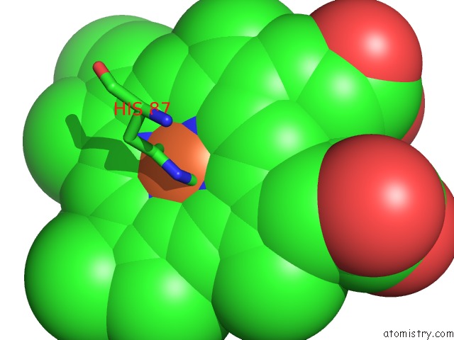
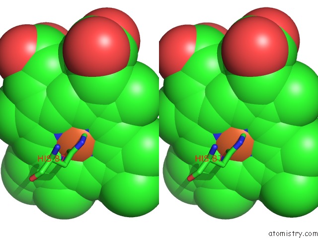
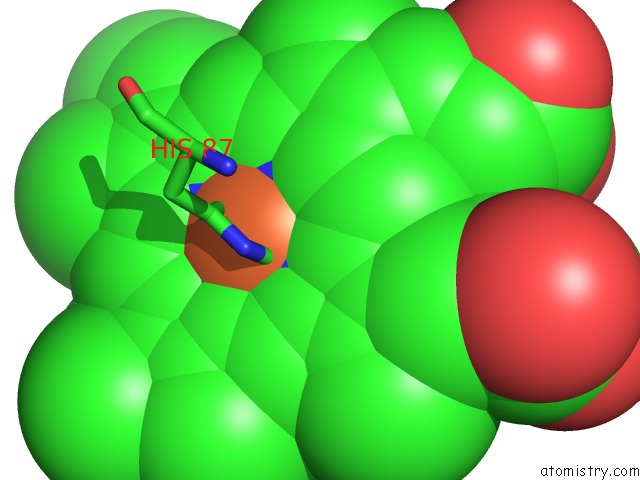
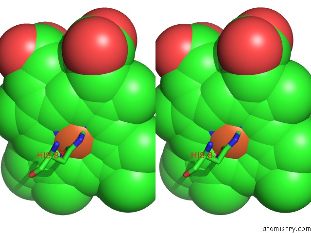
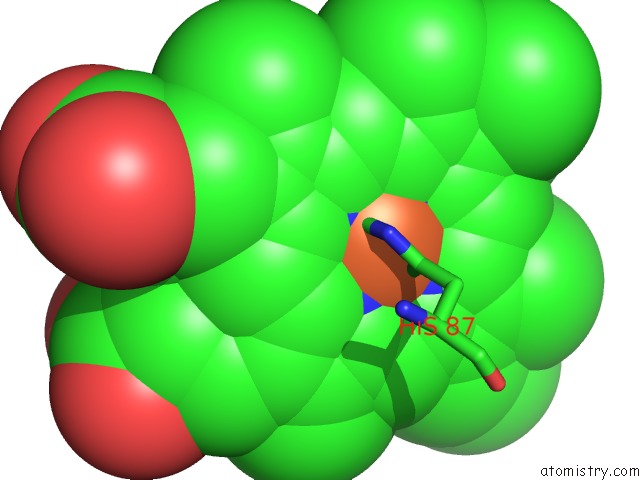
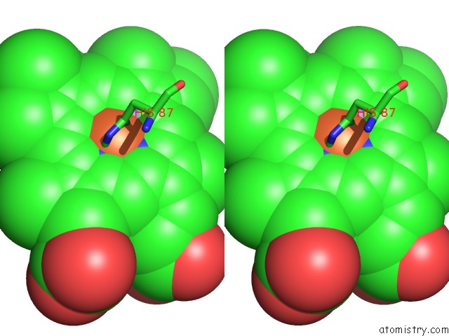
Iron binding site 1 out of 8 in 1hv4
Go back to
Iron binding site 1 out
of 8 in the Crystal Structure Analysis of Bar-Head Goose Hemoglobin (Deoxy Form)
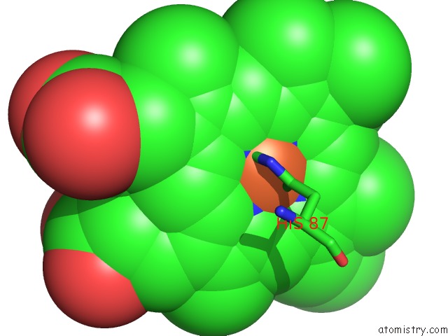
Mono view
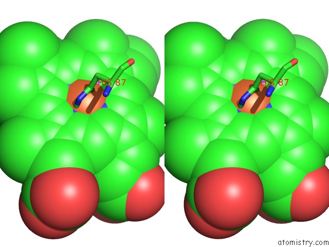
Stereo pair view
Mono view
Stereo pair view
A full contact list of Iron with other atoms in the Fe binding
site number 1 of Crystal Structure Analysis of Bar-Head Goose Hemoglobin (Deoxy Form) within 5.0Å range:
|
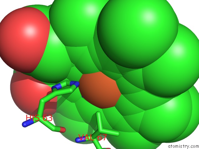
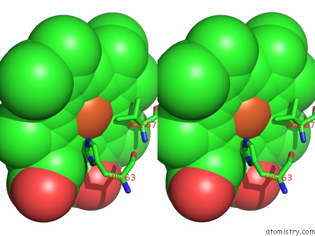
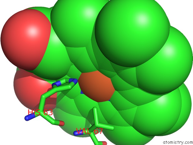
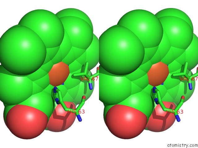
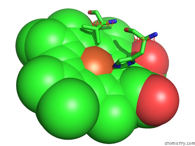
Iron binding site 2 out of 8 in 1hv4
Go back to
Iron binding site 2 out
of 8 in the Crystal Structure Analysis of Bar-Head Goose Hemoglobin (Deoxy Form)
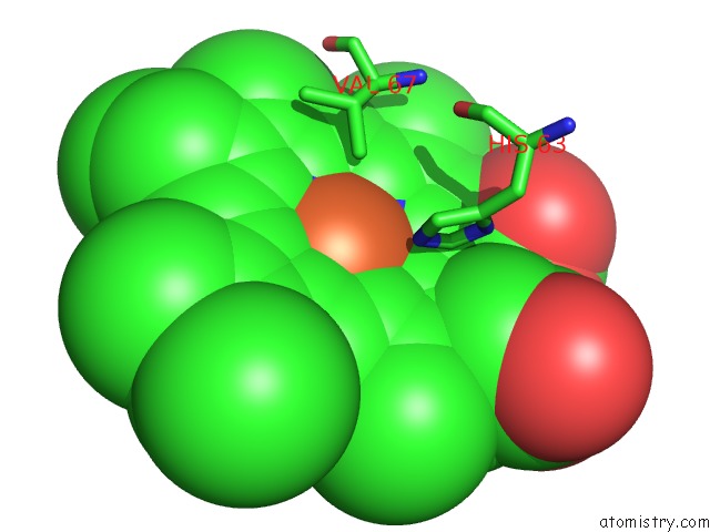
Mono view
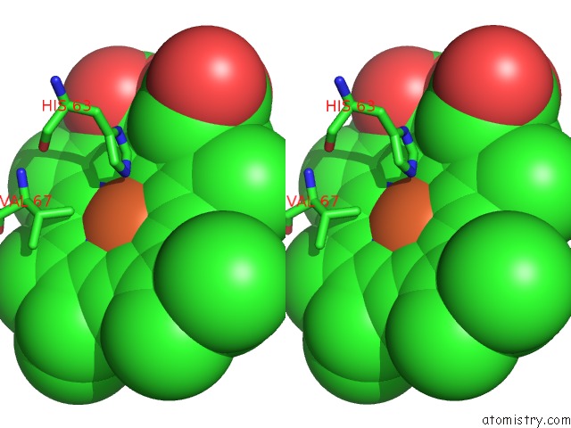
Stereo pair view
Mono view
Stereo pair view
A full contact list of Iron with other atoms in the Fe binding
site number 2 of Crystal Structure Analysis of Bar-Head Goose Hemoglobin (Deoxy Form) within 5.0Å range:
|
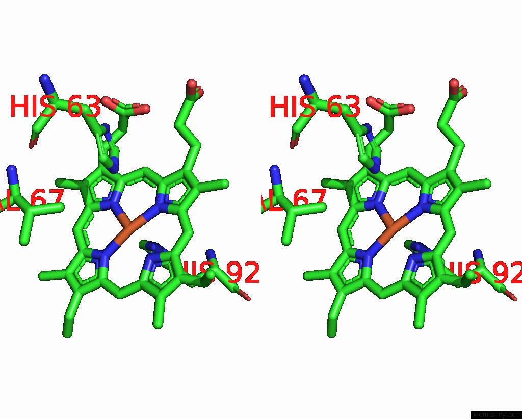
Iron binding site 3 out of 8 in 1hv4
Go back to
Iron binding site 3 out
of 8 in the Crystal Structure Analysis of Bar-Head Goose Hemoglobin (Deoxy Form)
Mono view
Stereo pair view
Mono view
Stereo pair view
A full contact list of Iron with other atoms in the Fe binding
site number 3 of Crystal Structure Analysis of Bar-Head Goose Hemoglobin (Deoxy Form) within 5.0Å range:
|
Iron binding site 4 out of 8 in 1hv4
Go back to
Iron binding site 4 out
of 8 in the Crystal Structure Analysis of Bar-Head Goose Hemoglobin (Deoxy Form)
Mono view
Stereo pair view
Mono view
Stereo pair view
A full contact list of Iron with other atoms in the Fe binding
site number 4 of Crystal Structure Analysis of Bar-Head Goose Hemoglobin (Deoxy Form) within 5.0Å range:
|
Iron binding site 5 out of 8 in 1hv4
Go back to
Iron binding site 5 out
of 8 in the Crystal Structure Analysis of Bar-Head Goose Hemoglobin (Deoxy Form)
Mono view
Stereo pair view
Mono view
Stereo pair view
A full contact list of Iron with other atoms in the Fe binding
site number 5 of Crystal Structure Analysis of Bar-Head Goose Hemoglobin (Deoxy Form) within 5.0Å range:
|
Iron binding site 6 out of 8 in 1hv4
Go back to
Iron binding site 6 out
of 8 in the Crystal Structure Analysis of Bar-Head Goose Hemoglobin (Deoxy Form)
Mono view
Stereo pair view
Mono view
Stereo pair view
A full contact list of Iron with other atoms in the Fe binding
site number 6 of Crystal Structure Analysis of Bar-Head Goose Hemoglobin (Deoxy Form) within 5.0Å range:
|
Iron binding site 7 out of 8 in 1hv4
Go back to
Iron binding site 7 out
of 8 in the Crystal Structure Analysis of Bar-Head Goose Hemoglobin (Deoxy Form)
Mono view
Stereo pair view
Mono view
Stereo pair view
A full contact list of Iron with other atoms in the Fe binding
site number 7 of Crystal Structure Analysis of Bar-Head Goose Hemoglobin (Deoxy Form) within 5.0Å range:
|
Iron binding site 8 out of 8 in 1hv4
Go back to
Iron binding site 8 out
of 8 in the Crystal Structure Analysis of Bar-Head Goose Hemoglobin (Deoxy Form)
Mono view
Stereo pair view
Mono view
Stereo pair view
A full contact list of Iron with other atoms in the Fe binding
site number 8 of Crystal Structure Analysis of Bar-Head Goose Hemoglobin (Deoxy Form) within 5.0Å range:
|
Reference:
Y.Liang,
Z.Hua,
X.Liang,
Q.Xu,
G.Lu.
The Crystal Structure of Bar-Headed Goose Hemoglobin in Deoxy Form: the Allosteric Mechanism of A Hemoglobin Species with High Oxygen Affinity. J.Mol.Biol. V. 313 123 2001.
ISSN: ISSN 0022-2836
PubMed: 11601851
DOI: 10.1006/JMBI.2001.5028
Page generated: Sat Aug 3 07:52:49 2024
ISSN: ISSN 0022-2836
PubMed: 11601851
DOI: 10.1006/JMBI.2001.5028
Last articles
Zn in 9J0NZn in 9J0O
Zn in 9J0P
Zn in 9FJX
Zn in 9EKB
Zn in 9C0F
Zn in 9CAH
Zn in 9CH0
Zn in 9CH3
Zn in 9CH1